


 Foundational characteristics of cancer include proliferation, angiogenesis, migration, evasion of apoptosis, and cellular immortality. Find key markers for these cellular processes and antibodies to detect them.
 The SUMOplot™ Analysis Program predicts and scores sumoylation sites in your protein. SUMOylation is a post-translational modification involved in various cellular processes, such as nuclear-cytosolic transport, transcriptional regulation, apoptosis, protein stability, response to stress, and progression through the cell cycle.
 The Autophagy Receptor Motif Plotter predicts and scores autophagy receptor binding sites in your protein. Identifying proteins connected to this pathway is critical to understanding the role of autophagy in physiological as well as pathological processes such as development, differentiation, neurodegenerative diseases, stress, infection, and cancer.
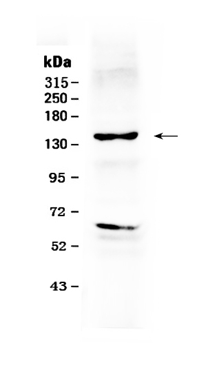
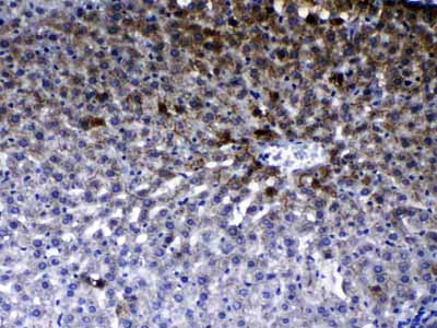
Anti-ErbB 2 Picoband Antibody

- SPECIFICATION
- CITATIONS
- PROTOCOLS
- BACKGROUND

Application
| WB, IHC-P |
|---|---|
| Primary Accession | P04626 |
| Host | Rabbit |
| Reactivity | Human, Rat |
| Clonality | Polyclonal |
| Format | Lyophilized |
| Description | Rabbit IgG polyclonal antibody for Receptor tyrosine-protein kinase erbB-2(ERBB2) detection. Tested with WB, IHC-P in Human;Rat. |
| Reconstitution | Add 0.2ml of distilled water will yield a concentration of 500ug/ml. |
| Gene ID | 2064 |
|---|---|
| Other Names | Receptor tyrosine-protein kinase erbB-2, 2.7.10.1, Metastatic lymph node gene 19 protein, MLN 19, Proto-oncogene Neu, Proto-oncogene c-ErbB-2, Tyrosine kinase-type cell surface receptor HER2, p185erbB2, CD340, ERBB2, HER2, MLN19, NEU, NGL |
| Calculated MW | 137910 MW KDa |
| Application Details | Immunohistochemistry(Paraffin-embedded Section), 0.5-1 µg/ml, Human, Rat, By Heat Western blot, 0.1-0.5 µg/ml, Human |
| Subcellular Localization | Isoform 1: Cell membrane; Single-pass type I membrane protein. Cytoplasm, perinuclear region. Nucleus. Translocation to the nucleus requires endocytosis, probably endosomal sorting and is mediated by importin beta-1/KPNB1. |
| Tissue Specificity | Expressed in a variety of tumor tissues including primary breast tumors and tumors from small bowel, esophagus, kidney and mouth. . |
| Protein Name | Receptor tyrosine-protein kinase erbB-2 |
| Contents | Each vial contains 5mg BSA, 0.9mg NaCl, 0.2mg Na2HPO4, 0.05mg NaN3. |
| Immunogen | A synthetic peptide corresponding to a sequence at the N-terminus of human ErbB 2 (29-64aa TDMKLRLPASPETHLDMLRHLYQGCQVVQGNLELTY), identical to the related mouse and rat sequences. |
| Purification | Immunogen affinity purified. |
| Cross Reactivity | No cross reactivity with other proteins |
| Storage | At -20˚C for one year. After r˚Constitution, at 4˚C for one month. It˚Can also be aliquotted and stored frozen at -20˚C for a longer time.Avoid repeated freezing and thawing. |
| Name | ERBB2 |
|---|---|
| Synonyms | HER2, MLN19, NEU, NGL |
| Function | Protein tyrosine kinase that is part of several cell surface receptor complexes, but that apparently needs a coreceptor for ligand binding. Essential component of a neuregulin-receptor complex, although neuregulins do not interact with it alone. GP30 is a potential ligand for this receptor. Regulates outgrowth and stabilization of peripheral microtubules (MTs). Upon ERBB2 activation, the MEMO1-RHOA-DIAPH1 signaling pathway elicits the phosphorylation and thus the inhibition of GSK3B at cell membrane. This prevents the phosphorylation of APC and CLASP2, allowing its association with the cell membrane. In turn, membrane-bound APC allows the localization of MACF1 to the cell membrane, which is required for microtubule capture and stabilization. |
| Cellular Location | Cell membrane; Single-pass type I membrane protein. Cell projection, ruffle membrane; Single-pass type I membrane protein. Note=Internalized from the cell membrane in response to EGF stimulation. [Isoform 2]: Cytoplasm. Nucleus. |
| Tissue Location | Expressed in a variety of tumor tissues including primary breast tumors and tumors from small bowel, esophagus, kidney and mouth. |

Thousands of laboratories across the world have published research that depended on the performance of antibodies from Abcepta to advance their research. Check out links to articles that cite our products in major peer-reviewed journals, organized by research category.
info@abcepta.com, and receive a free "I Love Antibodies" mug.

Provided below are standard protocols that you may find useful for product applications.
Background
Receptor tyrosine-protein kinase erbB-2, also known as CD340 (cluster of differentiation 340), proto-oncogene Neu, Erbb2 (rodent), or ERBB2 (human), is a protein that in humans is encoded by the ERBB2 gene. And it is also frequently called HER2 (from human epidermal growth factor receptor 2) or HER2/neu. This gene encodes a member of the epidermal growth factor (EGF) receptor family of receptor tyrosine kinases. This protein has no ligand binding domain of its own and therefore cannot bind growth factors. Amplification and/or overexpression of this gene has been reported in numerous cancers, including breast and ovarian tumors. Alternative splicing results in several additional transcript variants, some encoding different isoforms and others that have not been fully characterized.
If you have used an Abcepta product and would like to share how it has performed, please click on the "Submit Review" button and provide the requested information. Our staff will examine and post your review and contact you if needed.
If you have any additional inquiries please email technical services at tech@abcepta.com.
Ordering Information
Other Products
Shipping Information
