


 Foundational characteristics of cancer include proliferation, angiogenesis, migration, evasion of apoptosis, and cellular immortality. Find key markers for these cellular processes and antibodies to detect them.
 The SUMOplot™ Analysis Program predicts and scores sumoylation sites in your protein. SUMOylation is a post-translational modification involved in various cellular processes, such as nuclear-cytosolic transport, transcriptional regulation, apoptosis, protein stability, response to stress, and progression through the cell cycle.
 The Autophagy Receptor Motif Plotter predicts and scores autophagy receptor binding sites in your protein. Identifying proteins connected to this pathway is critical to understanding the role of autophagy in physiological as well as pathological processes such as development, differentiation, neurodegenerative diseases, stress, infection, and cancer.
Anti-HFE Picoband Antibody
- SPECIFICATION
- CITATIONS
- PROTOCOLS
- BACKGROUND

Application
| WB |
|---|---|
| Primary Accession | Q30201 |
| Host | Rabbit |
| Reactivity | Human |
| Clonality | Polyclonal |
| Format | Lyophilized |
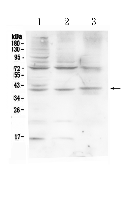
| Description | Rabbit IgG polyclonal antibody for Hereditary hemochromatosis protein(HFE) detection. Tested with WB in Human. |
| Reconstitution | Add 0.2ml of distilled water will yield a concentration of 500ug/ml. |
| Gene ID | 3077 |
|---|---|
| Other Names | Hereditary hemochromatosis protein, HLA-H, HFE, HLAH |
| Calculated MW | 40108 MW KDa |
| Application Details | Western blot, 0.1-0.5 µg/ml, Human |
| Subcellular Localization | Cell membrane ; Single-pass type I membrane protein . |
| Tissue Specificity | Expressed in all tissues tested except brain. |
| Protein Name | Hereditary hemochromatosis protein |
| Contents | Each vial contains 5mg BSA, 0.9mg NaCl, 0.2mg Na2HPO4, 0.05mg NaN3. |
| Immunogen | E.coli-derived human HFE recombinant protein (Position: Q82-R199). Human HFE shares 72.2% and 74.6% amino acid (aa) sequence identity with mouse and rat HFE, respectively. |
| Purification | Immunogen affinity purified. |
| Cross Reactivity | No cross reactivity with other proteins. |
| Storage | At -20˚C for one year. After r˚Constitution, at 4˚C for one month. It˚Can also be aliquotted and stored frozen at -20˚C for a longer time.Avoid repeated freezing and thawing. |
| Name | HFE |
|---|---|
| Synonyms | HLAH |
| Function | Binds to transferrin receptor (TFR) and reduces its affinity for iron-loaded transferrin. |
| Cellular Location | Cell membrane; Single-pass type I membrane protein |
| Tissue Location | Expressed in all tissues tested except brain. |

Thousands of laboratories across the world have published research that depended on the performance of antibodies from Abcepta to advance their research. Check out links to articles that cite our products in major peer-reviewed journals, organized by research category.
info@abcepta.com, and receive a free "I Love Antibodies" mug.

Provided below are standard protocols that you may find useful for product applications.
Background
Human hemochromatosis protein also known as the HFE protein is a protein which in humans is encoded by the HFE gene. The HFE gene is located on short arm of chromosome 6 at location 6p21.3. The protein encoded by this gene is a membrane protein that is similar to MHC class I-type proteins and associates with beta2-microglobulin (beta2M). It is thought that this protein functions to regulate iron absorption by regulating the interaction of the transferrin receptor with transferrin. The iron storage disorder, hereditary haemochromatosis, is a recessive genetic disorder that results from defects in this gene.
If you have used an Abcepta product and would like to share how it has performed, please click on the "Submit Review" button and provide the requested information. Our staff will examine and post your review and contact you if needed.
If you have any additional inquiries please email technical services at tech@abcepta.com.
Ordering Information
Other Products
Shipping Information
