


 Foundational characteristics of cancer include proliferation, angiogenesis, migration, evasion of apoptosis, and cellular immortality. Find key markers for these cellular processes and antibodies to detect them.
 The SUMOplot™ Analysis Program predicts and scores sumoylation sites in your protein. SUMOylation is a post-translational modification involved in various cellular processes, such as nuclear-cytosolic transport, transcriptional regulation, apoptosis, protein stability, response to stress, and progression through the cell cycle.
 The Autophagy Receptor Motif Plotter predicts and scores autophagy receptor binding sites in your protein. Identifying proteins connected to this pathway is critical to understanding the role of autophagy in physiological as well as pathological processes such as development, differentiation, neurodegenerative diseases, stress, infection, and cancer.
Anti-DVL1 Picoband Antibody
- SPECIFICATION
- CITATIONS
- PROTOCOLS
- BACKGROUND

Application
| WB, IHC-P |
|---|---|
| Primary Accession | O14640 |
| Host | Rabbit |
| Reactivity | Human, Rat |
| Clonality | Polyclonal |
| Format | Lyophilized |
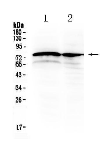
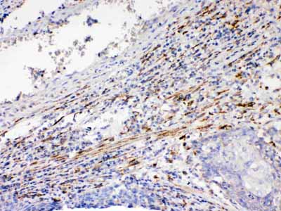
| Description | Rabbit IgG polyclonal antibody for Segment polarity protein dishevelled homolog DVL-1(DVL1) detection. Tested with WB, IHC-P in Human;Rat. |
| Reconstitution | Add 0.2ml of distilled water will yield a concentration of 500ug/ml. |
| Gene ID | 1855 |
|---|---|
| Other Names | Segment polarity protein dishevelled homolog DVL-1, Dishevelled-1, DSH homolog 1, DVL1 |
| Calculated MW | 75187 MW KDa |
| Application Details | Immunohistochemistry(Paraffin-embedded Section), 0.5-1 µg/ml, Human, By Heat Western blot, 0.1-0.5 µg/ml, Rat, Human |
| Subcellular Localization | Cell membrane ; Peripheral membrane protein ; Cytoplasmic side . Cytoplasm, cytosol . Cytoplasmic vesicle . Localizes at the cell membrane upon interaction with frizzled family members. . |
| Protein Name | Segment polarity protein dishevelled homolog DVL-1 |
| Contents | Each vial contains 5mg BSA, 0.9mg NaCl, 0.2mg Na2HPO4, 0.05mg NaN3. |
| Immunogen | A synthetic peptide corresponding to a sequence in the middle region of human DVL1 (401-438aa APQLEEAPLTVKSDMSAVVRVMQLPDSGLEIRDRMWLK), different from the related mouse and rat sequences by one amino acid. |
| Purification | Immunogen affinity purified. |
| Cross Reactivity | No cross reactivity with other proteins |
| Storage | At -20˚C for one year. After r˚Constitution, at 4˚C for one month. It˚Can also be aliquotted and stored frozen at -20˚C for a longer time.Avoid repeated freezing and thawing. |
| Name | DVL1 |
|---|---|
| Function | Participates in Wnt signaling by binding to the cytoplasmic C-terminus of frizzled family members and transducing the Wnt signal to down-stream effectors. Plays a role both in canonical and non-canonical Wnt signaling. Plays a role in the signal transduction pathways mediated by multiple Wnt genes. Required for LEF1 activation upon WNT1 and WNT3A signaling. DVL1 and PAK1 form a ternary complex with MUSK which is important for MUSK-dependent regulation of AChR clustering during the formation of the neuromuscular junction (NMJ). |
| Cellular Location | Cell membrane; Peripheral membrane protein; Cytoplasmic side. Cytoplasm, cytosol. Cytoplasmic vesicle Note=Localizes at the cell membrane upon interaction with frizzled family members. |

Thousands of laboratories across the world have published research that depended on the performance of antibodies from Abcepta to advance their research. Check out links to articles that cite our products in major peer-reviewed journals, organized by research category.
info@abcepta.com, and receive a free "I Love Antibodies" mug.

Provided below are standard protocols that you may find useful for product applications.
Background
Segment polarity protein dishevelled homolog DVL-1 is a protein that in humans is encoded by the DVL1 gene. DVL1, the human homolog of the Drosophila dishevelled gene (dsh) encodes a cytoplasmic phosphoprotein that regulates cell proliferation, acting as a transducer molecule for developmental processes, including segmentation and neuroblast specification. DVL1 is a candidate gene for neuroblastomatous transformation. The Schwartz-Jampel syndrome and Charcot-Marie-Tooth disease type 2A have been mapped to the same region as DVL1. The phenotypes of these diseases may be consistent with defects which might be expected from aberrant expression of a DVL gene during development.
If you have used an Abcepta product and would like to share how it has performed, please click on the "Submit Review" button and provide the requested information. Our staff will examine and post your review and contact you if needed.
If you have any additional inquiries please email technical services at tech@abcepta.com.
Ordering Information
Other Products
Shipping Information
