


 Foundational characteristics of cancer include proliferation, angiogenesis, migration, evasion of apoptosis, and cellular immortality. Find key markers for these cellular processes and antibodies to detect them.
 The SUMOplot™ Analysis Program predicts and scores sumoylation sites in your protein. SUMOylation is a post-translational modification involved in various cellular processes, such as nuclear-cytosolic transport, transcriptional regulation, apoptosis, protein stability, response to stress, and progression through the cell cycle.
 The Autophagy Receptor Motif Plotter predicts and scores autophagy receptor binding sites in your protein. Identifying proteins connected to this pathway is critical to understanding the role of autophagy in physiological as well as pathological processes such as development, differentiation, neurodegenerative diseases, stress, infection, and cancer.
Anti-PECAM-1/CD31 Antibody


- SPECIFICATION
- CITATIONS
- PROTOCOLS
- BACKGROUND

Application
| WB, IHC-P, IHC-F, FC, ICC |
|---|---|
| Primary Accession | P16284 |
| Host | Rabbit |
| Reactivity | Human |
| Clonality | Polyclonal |
| Format | Lyophilized |
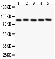
| Description | Rabbit IgG polyclonal antibody for Platelet endothelial cell adhesion molecule(PECAM1) detection. Tested with WB, IHC-P, IHC-F, ICC, FCM in Human. |
| Reconstitution | Add 0.2ml of distilled water will yield a concentration of 500ug/ml. |
| Gene ID | 5175 |
|---|---|
| Other Names | Platelet endothelial cell adhesion molecule, PECAM-1, EndoCAM, GPIIA', PECA1, CD31, PECAM1 |
| Calculated MW | 82536 MW KDa |
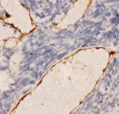
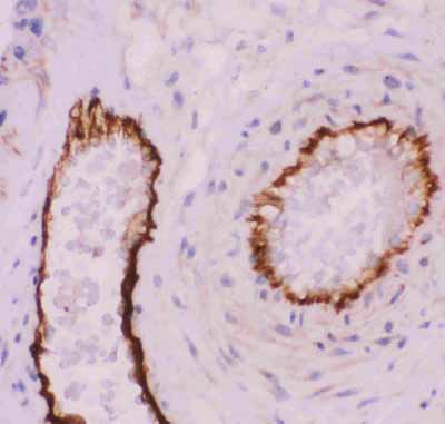
| Application Details | Immunohistochemistry(Paraffin-embedded Section), 0.5-1 µg/ml, By Heat Immunohistochemistry(Frozen Section), 0.5-1 µg/ml Immunocytochemistry, 0.5-1 µg/ml Western blot, 0.1-0.5 µg/ml Flow Cytometry, 1-3μg/1x106cells |
| Subcellular Localization | Isoform Long: Cell membrane; Single-pass type I membrane protein. Cell membrane; Lipid-anchor. Cell junction. Localizes to the lateral border recycling compartment (LBRC) and recycles from the LBRC to the junction in resting endothelial cells. |
| Tissue Specificity | Expressed on platelets and leukocytes and is primarily concentrated at the borders between endothelial cells. Isoform Long predominates in all tissues examined. Isoform Delta12 is detected only in trachea. Isoform Delta14-15 is only detected in lung. Isoform Delta14 is detected in all tissues examined with the strongest expression in heart. Isoform Delta15 is expressed in brain, testis, ovary, cell surface of platelets, human umbilical vein endothelial cells (HUVECs), Jurkat T-cell leukemia, human erythroleukemia (HEL) and U-937 histiocytic lymphoma cell lines (at protein level). . |
| Protein Name | Platelet endothelial cell adhesion molecule |
| Contents | Each vial contains 5mg BSA, 0.9mg NaCl, 0.2mg Na2HPO4, 0.05mg NaN3. |
| Immunogen | E.coli-derived human CD31 recombinant protein (Position: Q28-G382). Human CD31 shares 65% and 68% amino acid (aa) sequences identity with mouse and rat CD31, respectively. |
| Purification | Immunogen affinity purified. |
| Cross Reactivity | No cross reactivity with other proteins |
| Storage | At -20˚C for one year. After r˚Constitution, at 4˚C for one month. It˚Can also be aliquotted and stored frozen at -20˚C for a longer time.Avoid repeated freezing and thawing. |
| Sequence Similarities | Contains 6 Ig-like C2-type (immunoglobulin-like) domains. |
| Name | PECAM1 |
|---|---|
| Function | Cell adhesion molecule which is required for leukocyte transendothelial migration (TEM) under most inflammatory conditions (PubMed:19342684, PubMed:17580308). Tyr-690 plays a critical role in TEM and is required for efficient trafficking of PECAM1 to and from the lateral border recycling compartment (LBRC) and is also essential for the LBRC membrane to be targeted around migrating leukocytes (PubMed:19342684). Trans-homophilic interaction may play a role in endothelial cell-cell adhesion via cell junctions (PubMed:27958302). Heterophilic interaction with CD177 plays a role in transendothelial migration of neutrophils (PubMed:17580308). Homophilic ligation of PECAM1 prevents macrophage-mediated phagocytosis of neighboring viable leukocytes by transmitting a detachment signal (PubMed:12110892). Promotes macrophage-mediated phagocytosis of apoptotic leukocytes by tethering them to the phagocytic cells; PECAM1-mediated detachment signal appears to be disabled in apoptotic leukocytes (PubMed:12110892). Modulates bradykinin receptor BDKRB2 activation (PubMed:18672896). Regulates bradykinin- and hyperosmotic shock-induced ERK1/2 activation in endothelial cells (PubMed:18672896). Induces susceptibility to atherosclerosis (By similarity). |
| Cellular Location | Cell membrane; Single-pass type I membrane protein. Note=Cell surface expression on neutrophils is down-regulated upon fMLP or CXCL8/IL8- mediated stimulation. [Isoform Delta15]: Cell junction. Note=Localizes to the lateral border recycling compartment (LBRC) and recycles from the LBRC to the junction in resting endothelial cells |
| Tissue Location | Expressed on platelets and leukocytes and is primarily concentrated at the borders between endothelial cells (PubMed:18388311, PubMed:21464369). Expressed in human umbilical vein endothelial cells (HUVECs) (at protein level) (PubMed:19342684, PubMed:17580308). Expressed on neutrophils (at protein level) (PubMed:17580308). Isoform Long predominates in all tissues examined (PubMed:12433657). Isoform Delta12 is detected only in trachea (PubMed:12433657). Isoform Delta14-15 is only detected in lung (PubMed:12433657). Isoform Delta14 is detected in all tissues examined with the strongest expression in heart (PubMed:12433657). Isoform Delta15 is expressed in brain, testis, ovary, cell surface of platelets, human umbilical vein endothelial cells (HUVECs), Jurkat T- cell leukemia, human erythroleukemia (HEL) and U-937 histiocytic lymphoma cell lines (at protein level) (PubMed:12433657, PubMed:18388311). |

Thousands of laboratories across the world have published research that depended on the performance of antibodies from Abcepta to advance their research. Check out links to articles that cite our products in major peer-reviewed journals, organized by research category.
info@abcepta.com, and receive a free "I Love Antibodies" mug.

Provided below are standard protocols that you may find useful for product applications.
Background
CD31 also known as Platelet endothelial cell adhesion molecule (PECAM-1), is a protein that in human is encoded by the PECAM1 gene. Encoded protein is a member of the immunoglobulin superfamily, CD31 is mapped to 17q23.3. CD31 is found on the surface of platelets, monocytes, neutrophils, and some types of T-cells, and makes up a large portion of endothelial cell intercellular junctions. It is demonstrated that CD31 expression on human PBSCs may positively affect both neutrophil and platelet engraftment. Meanwhile, CD31 is involved in leukocyte migration and angiogenesis, which are key components of venous thrombus resolution.
If you have used an Abcepta product and would like to share how it has performed, please click on the "Submit Review" button and provide the requested information. Our staff will examine and post your review and contact you if needed.
If you have any additional inquiries please email technical services at tech@abcepta.com.
Ordering Information
Other Products
Shipping Information
