


 Foundational characteristics of cancer include proliferation, angiogenesis, migration, evasion of apoptosis, and cellular immortality. Find key markers for these cellular processes and antibodies to detect them.
 The SUMOplot™ Analysis Program predicts and scores sumoylation sites in your protein. SUMOylation is a post-translational modification involved in various cellular processes, such as nuclear-cytosolic transport, transcriptional regulation, apoptosis, protein stability, response to stress, and progression through the cell cycle.
 The Autophagy Receptor Motif Plotter predicts and scores autophagy receptor binding sites in your protein. Identifying proteins connected to this pathway is critical to understanding the role of autophagy in physiological as well as pathological processes such as development, differentiation, neurodegenerative diseases, stress, infection, and cancer.
Anti-CARD4 Picoband Antibody
- SPECIFICATION
- CITATIONS
- PROTOCOLS
- BACKGROUND

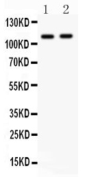
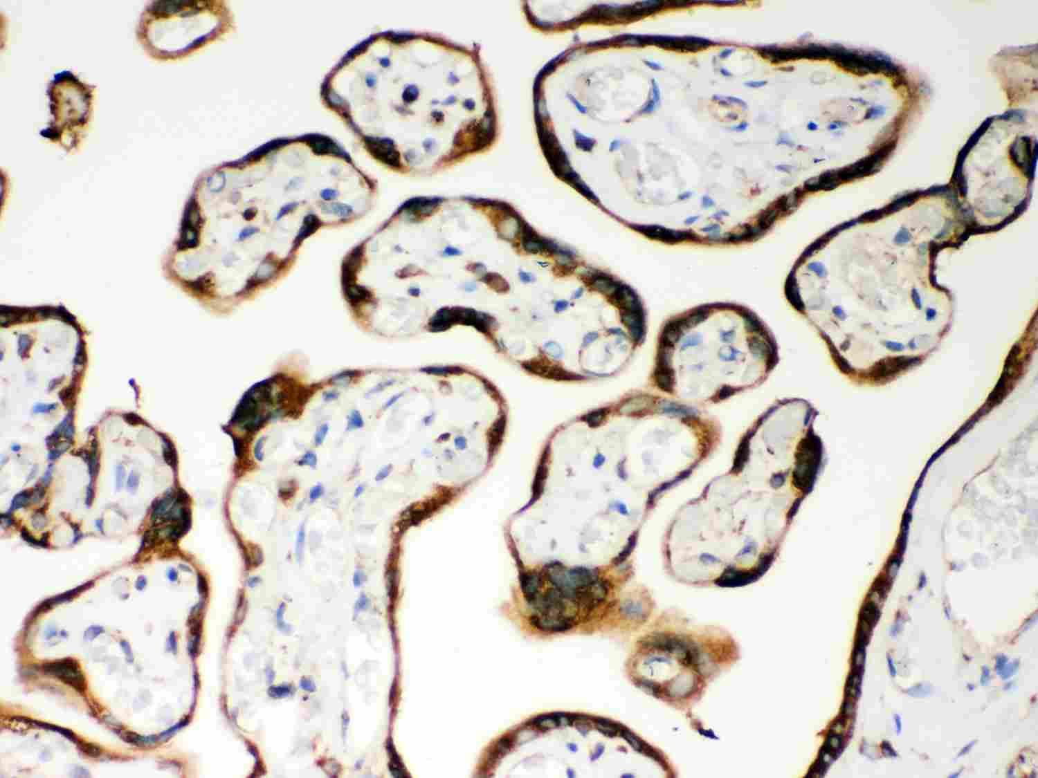
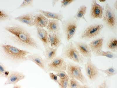
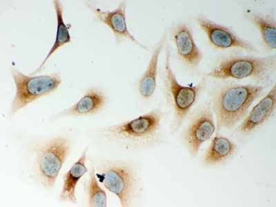
Application
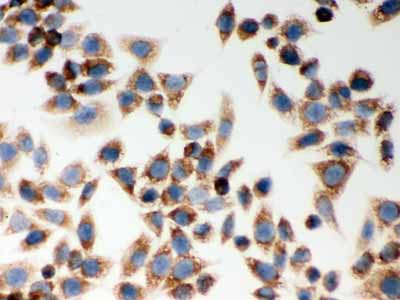
| WB, IHC-P, ICC |
|---|---|
| Primary Accession | Q9Y239 |
| Host | Rabbit |
| Reactivity | Human, Mouse, Rat |
| Clonality | Polyclonal |
| Format | Lyophilized |
| Description | Rabbit IgG polyclonal antibody for Nucleotide-binding oligomerization domain-containing protein 1(NOD1) detection. Tested with WB, IHC-P, ICC in Human;Mouse;Rat. |
| Reconstitution | Add 0.2ml of distilled water will yield a concentration of 500ug/ml. |
| Gene ID | 10392 |
|---|---|
| Other Names | Nucleotide-binding oligomerization domain-containing protein 1, Caspase recruitment domain-containing protein 4, NOD1, CARD4 |
| Calculated MW | 107691 MW KDa |
| Application Details | Immunohistochemistry(Paraffin-embedded Section), 0.5-1 µg/ml, By Heat Immunocytochemistry, 0.5-1 µg/ml Western blot, 0.1-0.5 µg/ml |
| Subcellular Localization | Cytoplasm. Cell membrane. Apical cell membrane. Basolateral cell membrane. Detected in the cytoplasm and at the cell membrane. Following bacterial infection, localizes to bacterial entry sites in the cell membrane. Recruited to the basolateral and apical membranes in polarized epithelial cells. |
| Tissue Specificity | Highly expressed in adult heart, skeletal muscle, pancreas, spleen and ovary. Also detected in placenta, lung, liver, kidney, thymus, testis, small intestine and colon. |
| Protein Name | Nucleotide-binding oligomerization domain-containing protein 1 |
| Contents | Each vial contains 5mg BSA, 0.9mg NaCl, 0.2mg Na2HPO4, 0.05mg NaN3. |
| Immunogen | E.coli-derived human CARD4 recombinant protein (Position: M1-M160). Human CARD4 shares 82% amino acid (aa) sequence identity with mouse CARD4. |
| Purification | Immunogen affinity purified. |
| Cross Reactivity | No cross reactivity with other proteins |
| Storage | At -20˚C for one year. After r˚Constitution, at 4˚C for one month. It˚Can also be aliquotted and stored frozen at -20˚C for a longer time.Avoid repeated freezing and thawing. |
| Sequence Similarities | Contains 1 CARD domain. |
| Name | NOD1 {ECO:0000303|PubMed:10329646, ECO:0000312|HGNC:HGNC:16390} |
|---|---|
| Function | Pattern recognition receptor (PRR) that detects bacterial peptidoglycan fragments and other danger signals and thus participates in both innate and adaptive immune responses (PubMed:11058605, PubMed:12796777, PubMed:12791997, PubMed:15044951, PubMed:16172124, PubMed:19043560, PubMed:22672233, PubMed:27099311). Specifically recognizes and binds gamma-D-glutamyl-meso-diaminopimelic acid (iE- DAP), a dipeptide present in peptidoglycan of Gram-negative bacteria (PubMed:12871942, PubMed:12796777, PubMed:12791997, PubMed:16211083, PubMed:16172124). Preferentially binds iE-DAP in tripeptide-containing muropeptides (MurNAc-TriDAP or TriDAP) (PubMed:16211083). Ligand binding triggers oligomerization that facilitates the binding and subsequent activation of the proximal adapter receptor-interacting RIPK2 (PubMed:12796777, PubMed:12791997, PubMed:17054981). Following recruitment, RIPK2 undergoes 'Met-1'- (linear) and 'Lys-63'-linked polyubiquitination by E3 ubiquitin-protein ligases XIAP, BIRC2, BIRC3 and the LUBAC complex, becoming a scaffolding protein for downstream effectors, triggering activation of the NF-kappa-B and MAP kinases signaling (PubMed:10880512, PubMed:12791997, PubMed:19043560). This in turn leads to the transcriptional activation of hundreds of genes involved in immune response (PubMed:10880512, PubMed:19043560). Also acts as a regulator of antiviral response elicited by dsRNA and the expression of RLR pathway members by targeting IFIH1 and TRAF3 to modulate the formation of IFIH1-MAVS and TRAF3-MAVS complexes leading to increased transcription of type I IFNs (PubMed:32169843). Also acts as a regulator of autophagy via its interaction with ATG16L1, possibly by recruiting ATG16L1 at the site of bacterial entry (By similarity). Besides recognizing pathogens, also involved in the endoplasmic reticulum stress response: acts by sensing and binding to the cytosolic metabolite sphingosine-1-phosphate generated in response to endoplasmic reticulum stress, initiating an inflammation process that leads to activation of the NF-kappa-B and MAP kinases signaling (PubMed:27007849, PubMed:33942347). In addition, plays a role in insulin trafficking in beta cells in a cell-autonomous manner (By similarity). Mechanistically, upon recognizing cognate ligands, NOD1 and RIPK2 localize to insulin vesicles where they recruit RAB1A to direct insulin trafficking through the cytoplasm (By similarity). |
| Cellular Location | Cell membrane; Lipid-anchor. Apical cell membrane. Basolateral cell membrane. Cytoplasm. Note=Detected in the cytoplasm and at the cell membrane (PubMed:31649195). Following bacterial infection, localizes to bacterial entry sites in the cell membrane (PubMed:31649195). Recruited to the basolateral and apical membranes in polarized epithelial cells (PubMed:19043560) |
| Tissue Location | Highly expressed in adult heart, skeletal muscle, pancreas, spleen and ovary (PubMed:10224040). Also detected in placenta, lung, liver, kidney, thymus, testis, small intestine and colon (PubMed:10224040). |

Thousands of laboratories across the world have published research that depended on the performance of antibodies from Abcepta to advance their research. Check out links to articles that cite our products in major peer-reviewed journals, organized by research category.
info@abcepta.com, and receive a free "I Love Antibodies" mug.

Provided below are standard protocols that you may find useful for product applications.
Background
Nucleotide-binding oligomerization domain-containing protein 1, also known as CARD4, is a protein receptor that in humans is encoded by the NOD1 gene. NOD1 is a member of NOD-like receptor protein family and is a close relative of NOD2. NOD1 is mapped to 7p14.3. It recognizes bacterial molecules and stimulates an immune reaction. NOD1 protein contains a caspase recruitment domain (CARD). This gene is an intracellular pattern recognition receptor, which is similar in structure to resistant proteins of plants, and mediates innate and acquired immunity by recognizing bacterial molecules containing D-glutamyl-meso-diaminopimelic acid (iE-DAP) moiety. What wore, it has been shown that NOD1 can sense cytosolic microbial products by monitoring the activation state of small Rho GTPases.
If you have used an Abcepta product and would like to share how it has performed, please click on the "Submit Review" button and provide the requested information. Our staff will examine and post your review and contact you if needed.
If you have any additional inquiries please email technical services at tech@abcepta.com.
Ordering Information
Other Products
Shipping Information
