


 Foundational characteristics of cancer include proliferation, angiogenesis, migration, evasion of apoptosis, and cellular immortality. Find key markers for these cellular processes and antibodies to detect them.
 The SUMOplot™ Analysis Program predicts and scores sumoylation sites in your protein. SUMOylation is a post-translational modification involved in various cellular processes, such as nuclear-cytosolic transport, transcriptional regulation, apoptosis, protein stability, response to stress, and progression through the cell cycle.
 The Autophagy Receptor Motif Plotter predicts and scores autophagy receptor binding sites in your protein. Identifying proteins connected to this pathway is critical to understanding the role of autophagy in physiological as well as pathological processes such as development, differentiation, neurodegenerative diseases, stress, infection, and cancer.
Anti-ERAB Antibody Antibody
- SPECIFICATION
- CITATIONS
- PROTOCOLS
- BACKGROUND

Application
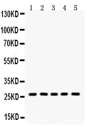
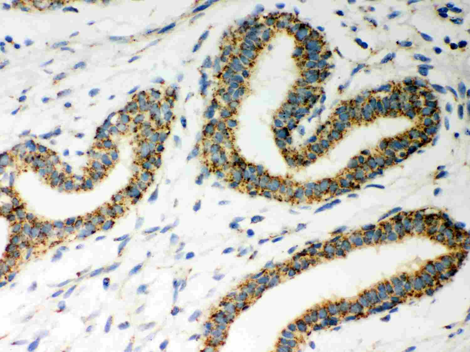
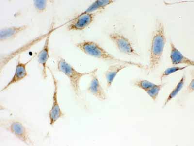
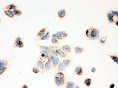
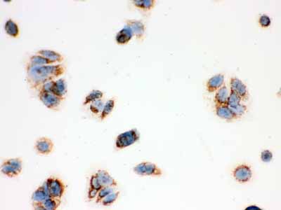
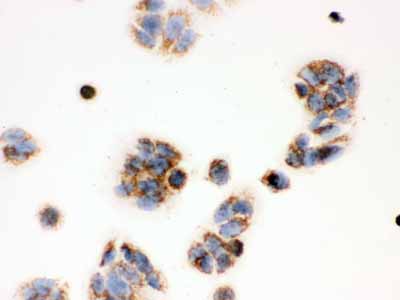
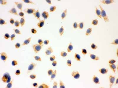
| WB, IHC-P, ICC |
|---|---|
| Primary Accession | Q99714 |
| Host | Rabbit |
| Reactivity | Human, Mouse, Rat |
| Clonality | Polyclonal |
| Format | Lyophilized |
| Description | Rabbit IgG polyclonal antibody for 3-hydroxyacyl-CoA dehydrogenase type-2(HSD17B10) detection. Tested with WB, IHC-P, ICC in Human;Mouse;Rat. |
| Reconstitution | Add 0.2ml of distilled water will yield a concentration of 500ug/ml. |
| Gene ID | 3028 |
|---|---|
| Other Names | 3-hydroxyacyl-CoA dehydrogenase type-2, 1.1.1.35, 17-beta-hydroxysteroid dehydrogenase 10, 17-beta-HSD 10, 1.1.1.51, 3-hydroxy-2-methylbutyryl-CoA dehydrogenase, 1.1.1.178, 3-hydroxyacyl-CoA dehydrogenase type II, Endoplasmic reticulum-associated amyloid beta-peptide-binding protein, Mitochondrial ribonuclease P protein 2, Mitochondrial RNase P protein 2, Short chain dehydrogenase/reductase family 5C member 1, Short-chain type dehydrogenase/reductase XH98G2, Type II HADH, HSD17B10, ERAB, HADH2, MRPP2, SCHAD, SDR5C1, XH98G2 |
| Calculated MW | 26923 MW KDa |
| Application Details | Immunohistochemistry(Paraffin-embedded Section), 0.5-1 µg/ml, By Heat Immunocytochemistry, 0.5-1 µg/ml Western blot, 0.1-0.5 µg/ml |
| Subcellular Localization | Mitochondrion . |
| Tissue Specificity | Ubiquitously expressed in normal tissues but is overexpressed in neurons affected in AD. . |
| Protein Name | 3-hydroxyacyl-CoA dehydrogenase type-2 |
| Contents | Each vial contains 5mg BSA, 0.9mg NaCl, 0.2mg Na2HPO4, 0.05mg NaN3. |
| Immunogen | E.coli-derived human ERAB recombinant protein (Position: E48-P261). Human ERAB shares 87% and 88% amino acid (aa) sequence identity with mouse and rat ERAB, respectively. |
| Purification | Immunogen affinity purified. |
| Cross Reactivity | No cross reactivity with other proteins |
| Storage | At -20˚C for one year. After r˚Constitution, at 4˚C for one month. It˚Can also be aliquotted and stored frozen at -20˚C for a longer time.Avoid repeated freezing and thawing. |
| Name | HSD17B10 |
|---|---|
| Synonyms | ERAB, HADH2, MRPP2, SCHAD, SDR5C1, XH98G |
| Function | Mitochondrial dehydrogenase involved in pathways of fatty acid, branched-chain amino acid and steroid metabolism (PubMed:9553139, PubMed:10600649, PubMed:12917011, PubMed:20077426, PubMed:18996107, PubMed:19706438, PubMed:25925575, PubMed:26950678, PubMed:28888424). Acts as (S)-3-hydroxyacyl-CoA dehydrogenase in mitochondrial fatty acid beta-oxidation, a major degradation pathway of fatty acids. Catalyzes the third step in the beta-oxidation cycle, namely the reversible conversion of (S)-3-hydroxyacyl-CoA to 3-ketoacyl-CoA. Preferentially accepts straight medium- and short-chain acyl-CoA substrates with highest efficiency for (3S)-hydroxybutanoyl-CoA (PubMed:9553139, PubMed:10600649, PubMed:12917011, PubMed:25925575, PubMed:26950678). Acts as 3-hydroxy-2-methylbutyryl-CoA dehydrogenase in branched-chain amino acid catabolic pathway. Catalyzes the oxidation of 3-hydroxy-2- methylbutanoyl-CoA into 2-methyl-3-oxobutanoyl-CoA, a step in isoleucine degradation pathway (PubMed:20077426, PubMed:18996107, PubMed:19706438). Has hydroxysteroid dehydrogenase activity toward steroid hormones and bile acids. Catalyzes the oxidation of 3alpha-, 17beta-, 20beta- and 21-hydroxysteroids and 7alpha- and 7beta-hydroxy bile acids (PubMed:10600649, PubMed:12917011). Oxidizes allopregnanolone/brexanolone at the 3alpha-hydroxyl group, which is known to be critical for the activation of gamma-aminobutyric acid receptors (GABAARs) chloride channel (PubMed:19706438, PubMed:28888424). Has phospholipase C-like activity toward cardiolipin and its oxidized species. Likely oxidizes the 2'-hydroxyl in the head group of cardiolipin to form a ketone intermediate that undergoes nucleophilic attack by water and fragments into diacylglycerol, dihydroxyacetone and orthophosphate. Has higher affinity for cardiolipin with oxidized fatty acids and may degrade these species during the oxidative stress response to protect cells from apoptosis (PubMed:26338420). By interacting with intracellular amyloid-beta, it may contribute to the neuronal dysfunction associated with Alzheimer disease (AD) (PubMed:9338779). Essential for structural and functional integrity of mitochondria (PubMed:20077426). |
| Cellular Location | Mitochondrion. Mitochondrion matrix, mitochondrion nucleoid |
| Tissue Location | Ubiquitously expressed in normal tissues but is overexpressed in neurons affected in AD. |

Thousands of laboratories across the world have published research that depended on the performance of antibodies from Abcepta to advance their research. Check out links to articles that cite our products in major peer-reviewed journals, organized by research category.
info@abcepta.com, and receive a free "I Love Antibodies" mug.

Provided below are standard protocols that you may find useful for product applications.
Background
ERAB is also known as HSD17B10 or HADH2. This gene encodes 3-hydroxyacyl-CoA dehydrogenase type II, a member of the short-chain dehydrogenase/reductase superfamily. The gene product is a mitochondrial protein that catalyzes the oxidation of a wide variety of fatty acids and steroids, and is a subunit of mitochondrial ribonuclease P, which is involved in tRNA maturation. The protein has been implicated in the development of Alzheimer disease, and mutations in the gene are the cause of 17beta-hydroxysteroid dehydrogenase type 10 (HSD10) deficiency. Several alternatively spliced transcript variants have been identified, but the full-length nature of only two transcript variants has been determined.
If you have used an Abcepta product and would like to share how it has performed, please click on the "Submit Review" button and provide the requested information. Our staff will examine and post your review and contact you if needed.
If you have any additional inquiries please email technical services at tech@abcepta.com.
Ordering Information
Other Products
Shipping Information
