


 Foundational characteristics of cancer include proliferation, angiogenesis, migration, evasion of apoptosis, and cellular immortality. Find key markers for these cellular processes and antibodies to detect them.
 The SUMOplot™ Analysis Program predicts and scores sumoylation sites in your protein. SUMOylation is a post-translational modification involved in various cellular processes, such as nuclear-cytosolic transport, transcriptional regulation, apoptosis, protein stability, response to stress, and progression through the cell cycle.
 The Autophagy Receptor Motif Plotter predicts and scores autophagy receptor binding sites in your protein. Identifying proteins connected to this pathway is critical to understanding the role of autophagy in physiological as well as pathological processes such as development, differentiation, neurodegenerative diseases, stress, infection, and cancer.
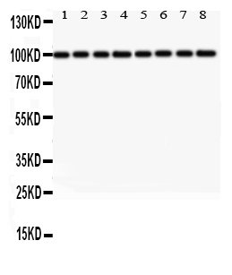
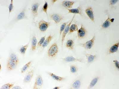
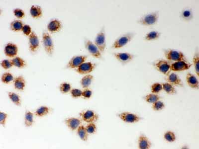
Anti-LYRIC Picoband Antibody
- SPECIFICATION
- CITATIONS
- PROTOCOLS
- BACKGROUND

Application
| WB, IHC-P, ICC |
|---|---|
| Primary Accession | Q86UE4 |
| Host | Rabbit |
| Reactivity | Human, Mouse, Rat |
| Clonality | Polyclonal |
| Format | Lyophilized |
| Description | Rabbit IgG polyclonal antibody for Protein LYRIC(MTDH) detection. Tested with WB, IHC-P, ICC in Human;Mouse;Rat. |
| Reconstitution | Add 0.2ml of distilled water will yield a concentration of 500ug/ml. |
| Gene ID | 92140 |
|---|---|
| Other Names | Protein LYRIC, 3D3/LYRIC, Astrocyte elevated gene-1 protein, AEG-1, Lysine-rich CEACAM1 co-isolated protein, Metadherin, Metastasis adhesion protein, MTDH, AEG1, LYRIC |
| Calculated MW | 63837 MW KDa |
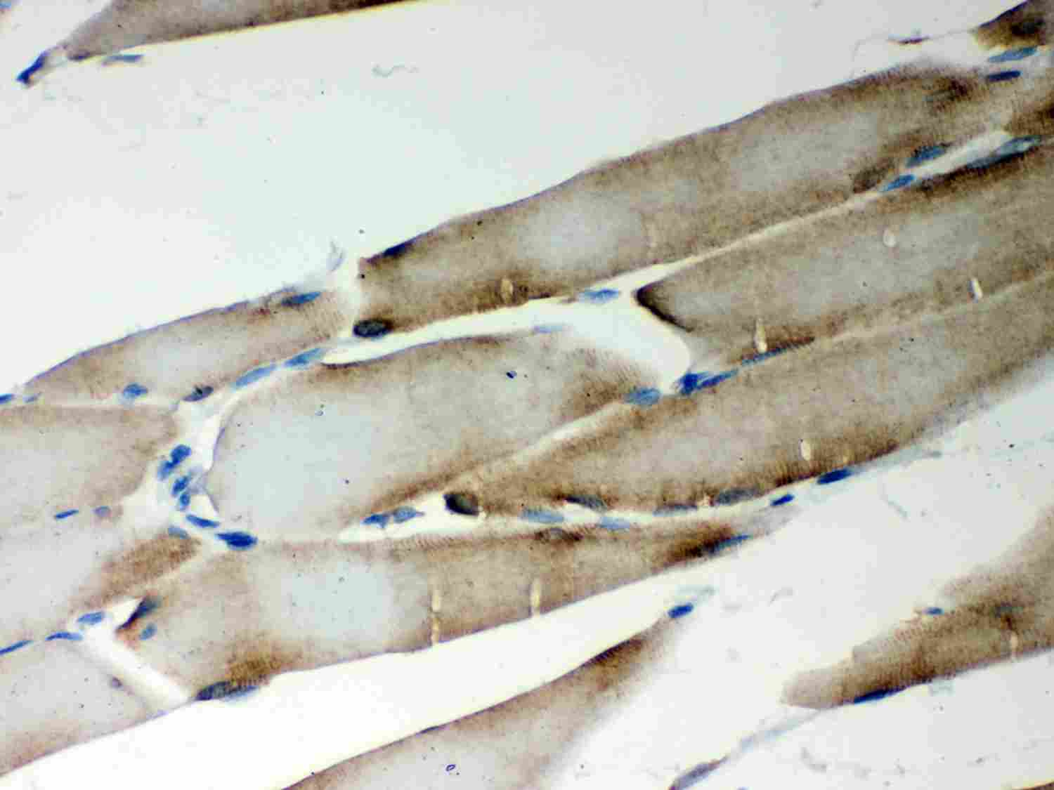
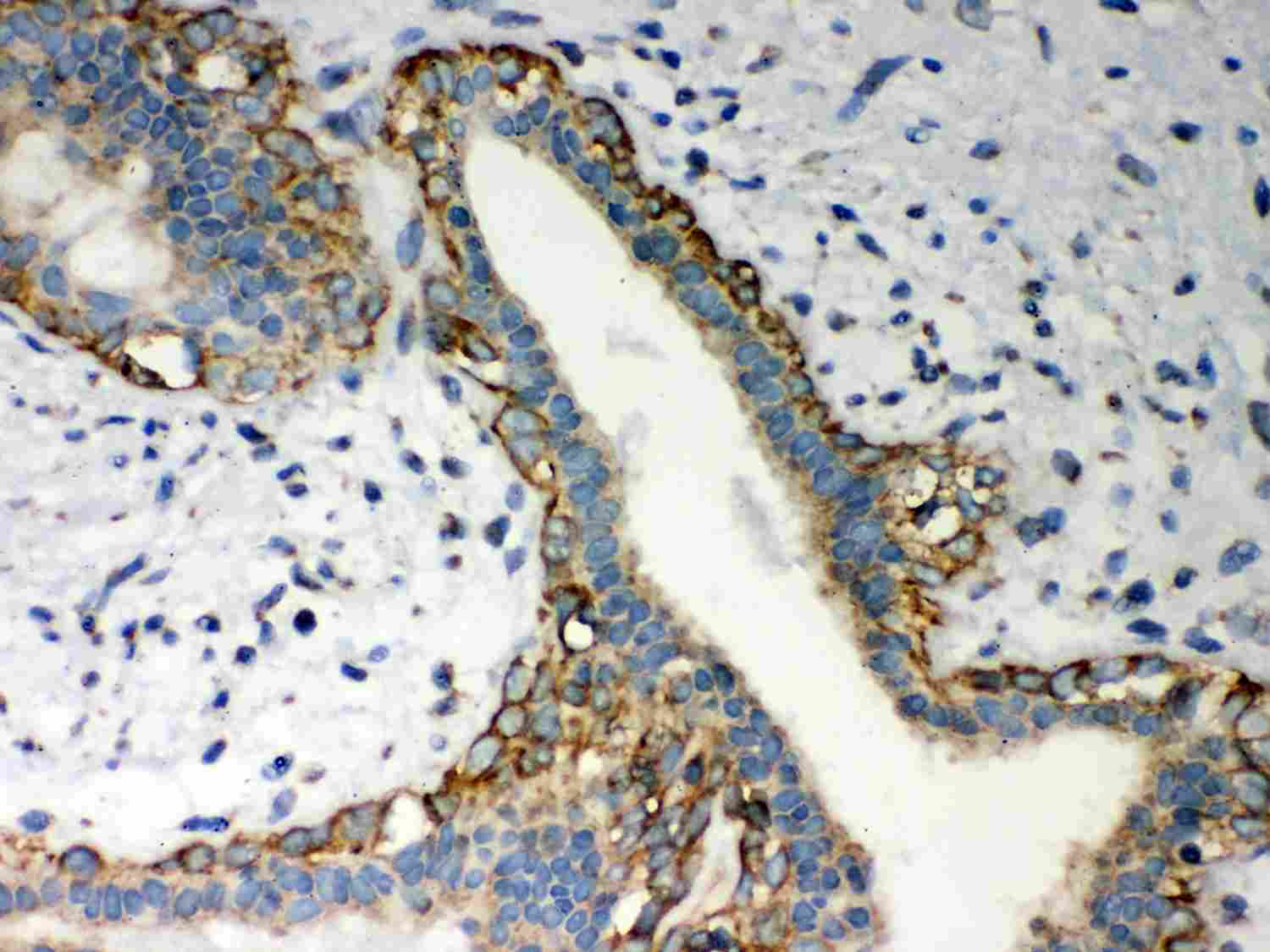
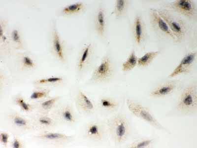
| Application Details | Immunohistochemistry(Paraffin-embedded Section), 0.5-1 µg/ml, By Heat Immunocytochemistry, 0.5-1 µg/ml Western blot, 0.1-0.5 µg/ml |
| Subcellular Localization | Endoplasmic reticulum membrane; Single-pass membrane protein. Nucleus membrane ; Single-pass membrane protein . Cell junction, tight junction . Nucleus, nucleolus . Cytoplasm, perinuclear region. In epithelial cells, recruited to tight junctions (TJ) during the maturation of the TJ complexes. A nucleolar staining may be due to nuclear targeting of an isoform lacking the transmembrane domain (By similarity). TNF-alpha causes translocation from the cytoplasm to the nucleus. . |
| Tissue Specificity | Widely expressed with highest levels in muscle-dominating organs such as skeletal muscle, heart, tongue and small intestine and in endocrine glands such as thyroid and adrenal gland. Overexpressed in various cancers including breast, brain, prostate, melanoma and glioblastoma multiforme. . |
| Protein Name | Protein LYRIC |
| Contents | Each vial contains 5mg BSA, 0.9mg NaCl, 0.2mg Na2HPO4, 0.05mg NaN3. |
| Immunogen | E.coli-derived human LYRIC recombinant protein (Position: D101-Q270). Human LYRIC shares 94% amino acid (aa) sequence identity with both mouse and rat LYRIC. |
| Purification | Immunogen affinity purified. |
| Cross Reactivity | No cross reactivity with other proteins |
| Storage | At -20˚C for one year. After r˚Constitution, at 4˚C for one month. It˚Can also be aliquotted and stored frozen at -20˚C for a longer time.Avoid repeated freezing and thawing. |
| Name | MTDH |
|---|---|
| Synonyms | AEG1, LYRIC |
| Function | Down-regulates SLC1A2/EAAT2 promoter activity when expressed ectopically. Activates the nuclear factor kappa-B (NF-kappa-B) transcription factor. Promotes anchorage-independent growth of immortalized melanocytes and astrocytes which is a key component in tumor cell expansion. Promotes lung metastasis and also has an effect on bone and brain metastasis, possibly by enhancing the seeding of tumor cells to the target organ endothelium. Induces chemoresistance. |
| Cellular Location | Endoplasmic reticulum membrane; Single-pass membrane protein. Nucleus membrane; Single-pass membrane protein. Cell junction, tight junction Nucleus, nucleolus. Cytoplasm, perinuclear region Note=In epithelial cells, recruited to tight junctions (TJ) during the maturation of the TJ complexes. A nucleolar staining may be due to nuclear targeting of an isoform lacking the transmembrane domain (By similarity). TNF-alpha causes translocation from the cytoplasm to the nucleus. |
| Tissue Location | Widely expressed with highest levels in muscle- dominating organs such as skeletal muscle, heart, tongue and small intestine and in endocrine glands such as thyroid and adrenal gland Overexpressed in various cancers including breast, brain, prostate, melanoma and glioblastoma multiforme. |

Thousands of laboratories across the world have published research that depended on the performance of antibodies from Abcepta to advance their research. Check out links to articles that cite our products in major peer-reviewed journals, organized by research category.
info@abcepta.com, and receive a free "I Love Antibodies" mug.

Provided below are standard protocols that you may find useful for product applications.
Background
MTDH (Metadherin), also known as protein LYRIC or astrocyte elevated gene-1 protein (AEG-1) is a protein that in humans is encoded by the MTDH gene. AEG-1 is involved in HIF-1alpha mediated angiogenesis. AEG-1 also interacts with SND1 and involved in RNA-induced silencing complex (RISC) and plays very important role in RISC and miRNA functions. AEG-1 induces an oncogene called Late SV40 factor (LSF/TFCP2) which is involved in thymidylate synthase (TS) induction and DNA biosynthesis synthesis. Late SV40 factor (LSF/TFCP2) enhances angiogenesis by transcriptionally up-regulating matrix metalloproteinase-9 (MMP9). AEG-1 acts as an oncogene in melanoma, malignant glioma, breast cancer and hepatocellular carcinoma. It is highly expressed in these cancers and helps in progression and development of these cancers. It is induced by c-Myc oncogene and plays very important role in anchorage independent growth of cancer cells.
If you have used an Abcepta product and would like to share how it has performed, please click on the "Submit Review" button and provide the requested information. Our staff will examine and post your review and contact you if needed.
If you have any additional inquiries please email technical services at tech@abcepta.com.
Ordering Information
Shipping Information
