


 Foundational characteristics of cancer include proliferation, angiogenesis, migration, evasion of apoptosis, and cellular immortality. Find key markers for these cellular processes and antibodies to detect them.
 The SUMOplot™ Analysis Program predicts and scores sumoylation sites in your protein. SUMOylation is a post-translational modification involved in various cellular processes, such as nuclear-cytosolic transport, transcriptional regulation, apoptosis, protein stability, response to stress, and progression through the cell cycle.
 The Autophagy Receptor Motif Plotter predicts and scores autophagy receptor binding sites in your protein. Identifying proteins connected to this pathway is critical to understanding the role of autophagy in physiological as well as pathological processes such as development, differentiation, neurodegenerative diseases, stress, infection, and cancer.
Anti-CD2 Picoband Antibody
- SPECIFICATION
- CITATIONS
- PROTOCOLS
- BACKGROUND

Application
| WB, IHC-P |
|---|---|
| Primary Accession | P08920 |
| Host | Rabbit |
| Reactivity | Mouse |
| Clonality | Polyclonal |
| Format | Lyophilized |
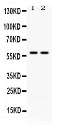
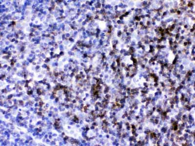
| Description | Rabbit IgG polyclonal antibody for T-cell surface antigen CD2(CD2) detection. Tested with WB, IHC-P in Mouse. |
| Reconstitution | Add 0.2ml of distilled water will yield a concentration of 500ug/ml. |
| Gene ID | 12481 |
|---|---|
| Other Names | T-cell surface antigen CD2, LFA-2, LFA-3 receptor, Lymphocyte antigen 37, Ly-37, T-cell surface antigen T11/Leu-5, CD2, Cd2, Ly-37 |
| Calculated MW | 38415 MW KDa |
| Application Details | Immunohistochemistry(Paraffin-embedded Section), 0.5-1 µg/ml, Mouse, By Heat Western blot, 0.1-0.5 µg/ml, Mouse |
| Subcellular Localization | Membrane; Single-pass type I membrane protein. |
| Protein Name | T-cell surface antigen CD2 |
| Contents | Each vial contains 5mg BSA, 0.9mg NaCl, 0.2mg Na2HPO4, 0.05mg NaN3. |
| Immunogen | E. coli-derived mouse CD2 recombinant protein (Position: R23-S203). Mouse CD2 shares 49.7% and 73.3% amino acid (aa) sequence identity with human and rat CD2, respectively. |
| Purification | Immunogen affinity purified. |
| Cross Reactivity | No cross reactivity with other proteins |
| Storage | At -20˚C for one year. After r˚Constitution, at 4˚C for one month. It˚Can also be aliquotted and stored frozen at -20˚C for a longer time.Avoid repeated freezing and thawing. |
| Name | Cd2 |
|---|---|
| Synonyms | Ly-37 |
| Function | CD2 interacts with lymphocyte function-associated antigen CD58 (LFA-3) and CD48/BCM1 to mediate adhesion between T-cells and other cell types. CD2 is implicated in the triggering of T-cells, the cytoplasmic domain is implicated in the signaling function. |
| Cellular Location | Cell membrane; Single-pass type I membrane protein |
| Tissue Location | Detected in thymus and spleen. |

Thousands of laboratories across the world have published research that depended on the performance of antibodies from Abcepta to advance their research. Check out links to articles that cite our products in major peer-reviewed journals, organized by research category.
info@abcepta.com, and receive a free "I Love Antibodies" mug.

Provided below are standard protocols that you may find useful for product applications.
Background
CD2 (cluster of differentiation 2) is a cell adhesion molecule found on the surface of T cells and natural killer (NK) cells. It has also been called T-cell surface antigen T11/Leu-5, LFA-2, LFA-3 receptor, erythrocyte receptor and rosette receptor. Monoclonal antibodies directed against CD2 inhibit the formation of rosettes with sheep erythrocytes, indicating that CD2 is the erythrocyte receptor or is closely associated with it. CD2 is one of the earliest T-cell markers, being present on more than 95% of thymocytes. Due to its structural characteristics, CD2 is a member of the immunoglobulin superfamily; it possesses two immunoglobulin-like domains in its extracellular portion. The localization of CD2 to 1p13 is established by in situ hybridization. CD2 interacts with other adhesion molecules, such as lymphocyte function-associated antigen-3 (LFA-3/CD58) in humans, or CD48 in rodents, which are expressed on the surfaces of other cells. With the use of transgenic mice, such an LCR is identified within the 3-prime flanking region of the human CD2 gene.
If you have used an Abcepta product and would like to share how it has performed, please click on the "Submit Review" button and provide the requested information. Our staff will examine and post your review and contact you if needed.
If you have any additional inquiries please email technical services at tech@abcepta.com.
Ordering Information
Other Products
Shipping Information
