


 Foundational characteristics of cancer include proliferation, angiogenesis, migration, evasion of apoptosis, and cellular immortality. Find key markers for these cellular processes and antibodies to detect them.
 The SUMOplot™ Analysis Program predicts and scores sumoylation sites in your protein. SUMOylation is a post-translational modification involved in various cellular processes, such as nuclear-cytosolic transport, transcriptional regulation, apoptosis, protein stability, response to stress, and progression through the cell cycle.
 The Autophagy Receptor Motif Plotter predicts and scores autophagy receptor binding sites in your protein. Identifying proteins connected to this pathway is critical to understanding the role of autophagy in physiological as well as pathological processes such as development, differentiation, neurodegenerative diseases, stress, infection, and cancer.
Anti-PD-L1/B7-H1 Picoband Antibody
- SPECIFICATION
- CITATIONS
- PROTOCOLS
- BACKGROUND

Application
| WB, E |
|---|---|
| Primary Accession | Q9EP73 |
| Host | Rabbit |
| Reactivity | Mouse |
| Clonality | Polyclonal |
| Format | Lyophilized |
| Description | Rabbit IgG polyclonal antibody for Programmed cell death 1 ligand 1(CD274) detection. Tested with WB, ELISA in Mouse. |
| Reconstitution | Add 0.2ml of distilled water will yield a concentration of 500ug/ml. |
| Gene ID | 60533 |
|---|---|
| Other Names | Programmed cell death 1 ligand 1, PD-L1, PDCD1 ligand 1, Programmed death ligand 1, B7 homolog 1, B7-H1, CD274, Cd274, B7h1, Pdcd1l1, Pdcd1lg1, Pdl1 |
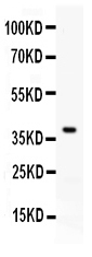
| Calculated MW | 32780 MW KDa |
| Application Details | ELISA , 0.1-0.5 µg/ml, Mouse, - Western blot, 0.1-0.5 µg/ml, Mouse |
| Subcellular Localization | Cell membrane ; Single-pass type I membrane protein . |
| Tissue Specificity | Highly expressed in the heart, thymus, skeletal muscle, and lung. Weakly expressed in the kidney, spleen, thyroid, and liver. Expressed on activated dendritic cells, B- cells and macrophages. Expressed in numerous tumor cells lines of lymphoid origin. . |
| Protein Name | Programmed cell death 1 ligand 1 |
| Contents | Each vial contains 5mg BSA, 0.9mg NaCl, 0.2mg Na2HPO4, 0.05mg NaN3. |
| Immunogen | E. coli-derived mouse PD-L1 recombinant protein (Position: F19-T238). Mouse PD-L1 shares 78.6% amino acid (aa) sequence identity with human PD-L1. |
| Purification | Immunogen affinity purified. |
| Cross Reactivity | No cross reactivity with other proteins. |
| Storage | At -20˚C for one year. After r˚Constitution, at 4˚C for one month. It˚Can also be aliquotted and stored frozen at -20˚C for a longer time.Avoid repeated freezing and thawing. |
| Name | Cd274 |
|---|---|
| Synonyms | B7h1, Pdcd1l1, Pdcd1lg1, Pdl1 |
| Function | Plays a critical role in induction and maintenance of immune tolerance to self (PubMed:11238124). As a ligand for the inhibitory receptor PDCD1/PD-1, modulates the activation threshold of T-cells and limits T-cell effector response (PubMed:11238124). Through a yet unknown activating receptor, may costimulate T-cell subsets that predominantly produce interleukin-10 (IL10) (PubMed:11015443, PubMed:12719480). |
| Cellular Location | Cell membrane {ECO:0000250|UniProtKB:Q9NZQ7}; Single-pass type I membrane protein. Early endosome membrane {ECO:0000250|UniProtKB:Q9NZQ7}; Single-pass type I membrane protein. Recycling endosome membrane {ECO:0000250|UniProtKB:Q9NZQ7}; Single-pass type I membrane protein |
| Tissue Location | Highly expressed in the heart, thymus, skeletal muscle, and lung. Weakly expressed in the kidney, spleen, thyroid, and liver. Expressed on activated dendritic cells, B-cells and macrophages Expressed in numerous tumor cells lines of lymphoid origin |

Thousands of laboratories across the world have published research that depended on the performance of antibodies from Abcepta to advance their research. Check out links to articles that cite our products in major peer-reviewed journals, organized by research category.
info@abcepta.com, and receive a free "I Love Antibodies" mug.

Provided below are standard protocols that you may find useful for product applications.
Background
Programmed death-ligand 1 (PD-L1), also known as CD274 or B7-H1, is a protein that in humans is encoded by the CD274 gene. It is mapped to 9p24.1. PD-L1 is a 40kDa type 1 transmembrane protein that has been speculated to play a major role in suppressing the immune system during particular events such as pregnancy, tissue allografts, autoimmune disease and other disease states such as hepatitis. It has been concluded that upregulation of PD-L1 on tumor MDCs downregulates T-cell immunity and that PD-L1 blockade may represent an approach for cancer immunotherapy. Additionally, PD-L1 can provide positive costimulatory signals for innate and adaptive immunity and for protection against intracellular bacterial infection. What’s more, it has been found that PD1/PDL1 pathway may be a good target for restoring antitumor immunity in ovarian cancer.
If you have used an Abcepta product and would like to share how it has performed, please click on the "Submit Review" button and provide the requested information. Our staff will examine and post your review and contact you if needed.
If you have any additional inquiries please email technical services at tech@abcepta.com.
Ordering Information
Other Products
Shipping Information
