


 Foundational characteristics of cancer include proliferation, angiogenesis, migration, evasion of apoptosis, and cellular immortality. Find key markers for these cellular processes and antibodies to detect them.
 The SUMOplot™ Analysis Program predicts and scores sumoylation sites in your protein. SUMOylation is a post-translational modification involved in various cellular processes, such as nuclear-cytosolic transport, transcriptional regulation, apoptosis, protein stability, response to stress, and progression through the cell cycle.
 The Autophagy Receptor Motif Plotter predicts and scores autophagy receptor binding sites in your protein. Identifying proteins connected to this pathway is critical to understanding the role of autophagy in physiological as well as pathological processes such as development, differentiation, neurodegenerative diseases, stress, infection, and cancer.
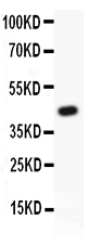
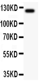
Anti-TIE2 Antibody
- SPECIFICATION
- CITATIONS
- PROTOCOLS
- BACKGROUND

Application
| WB |
|---|---|
| Primary Accession | Q02763 |
| Host | Rabbit |
| Reactivity | Human |
| Clonality | Polyclonal |
| Format | Lyophilized |
| Description | Rabbit IgG polyclonal antibody for Angiopoietin-1 receptor(TEK) detection. Tested with WB in Human. |
| Reconstitution | Add 0.2ml of distilled water will yield a concentration of 500ug/ml. |
| Gene ID | 7010 |
|---|---|
| Other Names | Angiopoietin-1 receptor, 2.7.10.1, Endothelial tyrosine kinase, Tunica interna endothelial cell kinase, Tyrosine kinase with Ig and EGF homology domains-2, Tyrosine-protein kinase receptor TEK, Tyrosine-protein kinase receptor TIE-2, hTIE2, p140 TEK, CD202b, TEK, TIE2, VMCM, VMCM1 |
| Calculated MW | 125830 MW KDa |
| Application Details | Western blot, 0.1-0.5 µg/ml, Human |
| Subcellular Localization | Cell membrane; Single-pass type I membrane protein. Cell junction. Cell junction, focal adhesion. Cytoplasm, cytoskeleton. Secreted. Recruited to cell-cell contacts in quiescent endothelial cells. Colocalizes with the actin cytoskeleton and at actin stress fibers during cell spreading. Recruited to the lower surface of migrating cells, especially the rear end of the cell. Proteolytic processing gives rise to a soluble extracellular domain that is secreted. |
| Tissue Specificity | Detected in umbilical vein endothelial cells. Proteolytic processing gives rise to a soluble extracellular domain that is detected in blood plasma (at protein level). Predominantly expressed in endothelial cells and their progenitors, the angioblasts. Has been directly found in placenta and lung, with a lower level in umbilical vein endothelial cells, brain and kidney. . |
| Protein Name | Angiopoietin-1 receptor |
| Contents | Each vial contains 5mg BSA, 0.9mg NaCl, 0.2mg Na2HPO4, 0.05mg NaN3. |
| Immunogen | E.coli-derived human TIE2 recombinant protein (Position: Q641-I830). Human TIE2 shares 91% amino acid (aa) sequence identity with mouse TIE2. |
| Purification | Immunogen affinity purified. |
| Cross Reactivity | No cross reactivity with other proteins |
| Storage | At -20˚C for one year. After r˚Constitution, at 4˚C for one month. It˚Can also be aliquotted and stored frozen at -20˚C for a longer time.Avoid repeated freezing and thawing. |
| Sequence Similarities | Belongs to the protein kinase superfamily. Tyr protein kinase family. Tie subfamily. |
| Name | TEK (HGNC:11724) |
|---|---|
| Function | Tyrosine-protein kinase that acts as a cell-surface receptor for ANGPT1, ANGPT2 and ANGPT4 and regulates angiogenesis, endothelial cell survival, proliferation, migration, adhesion and cell spreading, reorganization of the actin cytoskeleton, but also maintenance of vascular quiescence. Has anti-inflammatory effects by preventing the leakage of pro-inflammatory plasma proteins and leukocytes from blood vessels. Required for normal angiogenesis and heart development during embryogenesis. Required for post-natal hematopoiesis. After birth, activates or inhibits angiogenesis, depending on the context. Inhibits angiogenesis and promotes vascular stability in quiescent vessels, where endothelial cells have tight contacts. In quiescent vessels, ANGPT1 oligomers recruit TEK to cell-cell contacts, forming complexes with TEK molecules from adjoining cells, and this leads to preferential activation of phosphatidylinositol 3-kinase and the AKT1 signaling cascades. In migrating endothelial cells that lack cell-cell adhesions, ANGT1 recruits TEK to contacts with the extracellular matrix, leading to the formation of focal adhesion complexes, activation of PTK2/FAK and of the downstream kinases MAPK1/ERK2 and MAPK3/ERK1, and ultimately to the stimulation of sprouting angiogenesis. ANGPT1 signaling triggers receptor dimerization and autophosphorylation at specific tyrosine residues that then serve as binding sites for scaffold proteins and effectors. Signaling is modulated by ANGPT2 that has lower affinity for TEK, can promote TEK autophosphorylation in the absence of ANGPT1, but inhibits ANGPT1-mediated signaling by competing for the same binding site. Signaling is also modulated by formation of heterodimers with TIE1, and by proteolytic processing that gives rise to a soluble TEK extracellular domain. The soluble extracellular domain modulates signaling by functioning as decoy receptor for angiopoietins. TEK phosphorylates DOK2, GRB7, GRB14, PIK3R1; SHC1 and TIE1. |
| Cellular Location | Cell membrane; Single-pass type I membrane protein. Cell junction. Cell junction, focal adhesion. Cytoplasm, cytoskeleton. Secreted. Note=Recruited to cell-cell contacts in quiescent endothelial cells (PubMed:18425120, PubMed:18425119) Colocalizes with the actin cytoskeleton and at actin stress fibers during cell spreading. Recruited to the lower surface of migrating cells, especially the rear end of the cell. Proteolytic processing gives rise to a soluble extracellular domain that is secreted (PubMed:11806244). |
| Tissue Location | Detected in umbilical vein endothelial cells. Proteolytic processing gives rise to a soluble extracellular domain that is detected in blood plasma (at protein level). Predominantly expressed in endothelial cells and their progenitors, the angioblasts Has been directly found in placenta and lung, with a lower level in umbilical vein endothelial cells, brain and kidney |

Thousands of laboratories across the world have published research that depended on the performance of antibodies from Abcepta to advance their research. Check out links to articles that cite our products in major peer-reviewed journals, organized by research category.
info@abcepta.com, and receive a free "I Love Antibodies" mug.

Provided below are standard protocols that you may find useful for product applications.
Background
TIE2, also known as TEK tyrosine kinase, TIE2 gene is mapped to 9p21.2. This gene encodes a receptor that belongs to the protein tyrosine kinase Tie2 family. The encoded protein possesses a unique extracellular region that contains two immunoglobulin-like domains, three epidermal growth factor (EGF)-like domains and three fibronectin type III repeats. The ligand angiopoietin-1 binds to this receptor and mediates a signaling pathway that functions in embryonic vascular development. Immunoblotting showed that TIE2 expression was increased by thyroid-stimulating hormone and agents that increased intracellular cAMP. HSCs expressing the receptor tyrosine kinase TIE2 are quiescent and antiapoptotic and comprise a side population of HSCs that adhere to osteoblasts in the bone marrow niche.
If you have used an Abcepta product and would like to share how it has performed, please click on the "Submit Review" button and provide the requested information. Our staff will examine and post your review and contact you if needed.
If you have any additional inquiries please email technical services at tech@abcepta.com.
Ordering Information
Other Products
Shipping Information
