


 Foundational characteristics of cancer include proliferation, angiogenesis, migration, evasion of apoptosis, and cellular immortality. Find key markers for these cellular processes and antibodies to detect them.
 The SUMOplot™ Analysis Program predicts and scores sumoylation sites in your protein. SUMOylation is a post-translational modification involved in various cellular processes, such as nuclear-cytosolic transport, transcriptional regulation, apoptosis, protein stability, response to stress, and progression through the cell cycle.
 The Autophagy Receptor Motif Plotter predicts and scores autophagy receptor binding sites in your protein. Identifying proteins connected to this pathway is critical to understanding the role of autophagy in physiological as well as pathological processes such as development, differentiation, neurodegenerative diseases, stress, infection, and cancer.
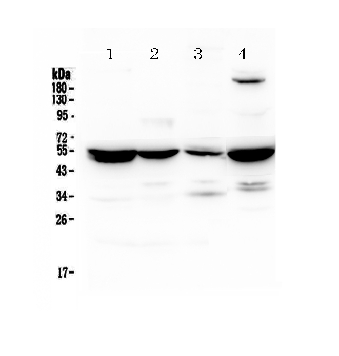
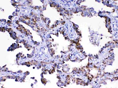
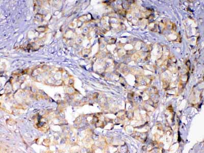
Anti-Adenylosuccinate Lyase Picoband Antibody
- SPECIFICATION
- CITATIONS
- PROTOCOLS
- BACKGROUND

Application
| WB, IHC-P |
|---|---|
| Primary Accession | P04424 |
| Host | Rabbit |
| Reactivity | Human, Mouse, Rat |
| Clonality | Polyclonal |
| Format | Lyophilized |
| Description | Rabbit IgG polyclonal antibody for Adenylosuccinate Lyase detection. Tested with WB, IHC-P in Human;Mouse;Rat. |
| Reconstitution | Add 0.2ml of distilled water will yield a concentration of 500ug/ml. |
| Gene ID | 435 |
|---|---|
| Other Names | Argininosuccinate lyase, ASAL, 4.3.2.1, Arginosuccinase, ASL |
| Calculated MW | 51658 MW KDa |
| Application Details | Western blot, 0.1-0.5 µg/ml Immunohistochemistry(Paraffin-embedded Section), 0.5-1 µg/ml |
| Contents | Each vial contains 5mg BSA, 0.9mg NaCl, 0.2mg Na2HPO4, 0.05mg NaN3. |
| Immunogen | A synthetic peptide corresponding to a sequence of human Adenylosuccinate Lyase (YTHLQRAQPIRWSHWILSHAVALTRDSERLLEVRKRIN). |
| Purification | Immunogen affinity purified. |
| Cross Reactivity | No cross reactivity with other proteins. |
| Storage | At -20˚C for one year. After r˚Constitution, at 4˚C for one month. It˚Can also be aliquotted and stored frozen at -20˚C for a longer time. Avoid repeated freezing and thawing. |
| Name | ASL |
|---|---|
| Function | Catalyzes the reversible cleavage of L-argininosuccinate to fumarate and L-arginine, an intermediate step reaction in the urea cycle mostly providing for hepatic nitrogen detoxification into excretable urea as well as de novo L-arginine synthesis in nonhepatic tissues (PubMed:11747433, PubMed:11747432, PubMed:9045711, PubMed:22081021, PubMed:2263616). Essential regulator of intracellular and extracellular L-arginine pools. As part of citrulline-nitric oxide cycle, forms tissue-specific multiprotein complexes with argininosuccinate synthase ASS1, transport protein SLC7A1 and nitric oxide synthase NOS1, NOS2 or NOS3, allowing for cell-autonomous L- arginine synthesis while channeling extracellular L-arginine to nitric oxide synthesis pathway (PubMed:22081021). |

Thousands of laboratories across the world have published research that depended on the performance of antibodies from Abcepta to advance their research. Check out links to articles that cite our products in major peer-reviewed journals, organized by research category.
info@abcepta.com, and receive a free "I Love Antibodies" mug.

Provided below are standard protocols that you may find useful for product applications.
Background
ASL (argininosuccinatelyase, also known as argininosuccinase) is an enzyme that catalyzes the reversible breakdown of argininosuccinate (ASA) producing the amino acid arginine and dicarboxylic acid fumarate. Located in liver cytosol, ASL is the fourth enzyme of the urea cycle and involved in the biosynthesis of arginine in all species and the production of urea in ureotelic species. Mutations in ASL, resulting low activity of the enzyme, increase levels of urea in the body and result in various side effects. The ASL gene is located on chromosome 7 between the centromere (junction of the long and short arm) and the long (q) arm at position 11.2, from base pair 64,984,963 to base pair 65,002,090.
If you have used an Abcepta product and would like to share how it has performed, please click on the "Submit Review" button and provide the requested information. Our staff will examine and post your review and contact you if needed.
If you have any additional inquiries please email technical services at tech@abcepta.com.
Ordering Information
Other Products
Shipping Information
