


 Foundational characteristics of cancer include proliferation, angiogenesis, migration, evasion of apoptosis, and cellular immortality. Find key markers for these cellular processes and antibodies to detect them.
 The SUMOplot™ Analysis Program predicts and scores sumoylation sites in your protein. SUMOylation is a post-translational modification involved in various cellular processes, such as nuclear-cytosolic transport, transcriptional regulation, apoptosis, protein stability, response to stress, and progression through the cell cycle.
 The Autophagy Receptor Motif Plotter predicts and scores autophagy receptor binding sites in your protein. Identifying proteins connected to this pathway is critical to understanding the role of autophagy in physiological as well as pathological processes such as development, differentiation, neurodegenerative diseases, stress, infection, and cancer.
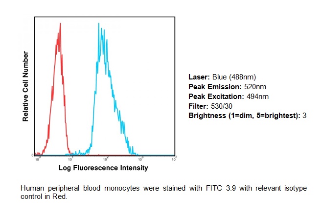
CD11c FITC Monoclonal Antibody (Clone 3.9)
Mouse Monoclonal Antibody
- SPECIFICATION
- CITATIONS
- PROTOCOLS
- BACKGROUND

Application
| FC |
|---|---|
| Primary Accession | P20702 |
| Reactivity | Human |
| Host | Mouse |
| Clonality | Monoclonal |
| Isotype | Mouse IgG1, Kappa |
| Clone Names | 3.9 |
| Calculated MW | 127829 Da |
| Gene ID | 3687 |
|---|---|
| Positive Control | FACS: Human PMBCs |
| Application & Usage | Flow (Cell Surface): 5 µl/1x10^6 cells, Volume per test: 5 µl (1 µg). |
| Other Names | CD11c |
| Target/Specificity | CD11c |
| Antibody Form | Liquid |
| Appearance | Colorless liquid |
| Formulation | Phosphate-buffered aqueous solution pH 7.2, ≤0.09% Sodium azide, may contain carrier protein/stabilizer. |
| Handling | The antibody solution should be gently mixed before use. |
| Reconstitution & Storage | 4°C |
| Background Descriptions | |
| Precautions | CD11c FITC Monoclonal Antibody (Clone 3.9) is for research use only and not for use in diagnostic or therapeutic procedures. |
| Name | ITGAX |
|---|---|
| Synonyms | CD11C |
| Function | Integrin alpha-X/beta-2 is a receptor for fibrinogen. It recognizes the sequence G-P-R in fibrinogen. It mediates cell-cell interaction during inflammatory responses. It is especially important in monocyte adhesion and chemotaxis. |
| Cellular Location | Membrane; Single-pass type I membrane protein. |
| Tissue Location | Predominantly expressed in monocytes and granulocytes |

Thousands of laboratories across the world have published research that depended on the performance of antibodies from Abcepta to advance their research. Check out links to articles that cite our products in major peer-reviewed journals, organized by research category.
info@abcepta.com, and receive a free "I Love Antibodies" mug.

Provided below are standard protocols that you may find useful for product applications.
Background
CD3 (T3), a complex T cell marker, is known to associate noncovalently with the a/b or g/z heterodimer of the T cell antigen receptor (TCR) to form the most complex transmembrane (TM) receptor structures. CD3 is specially engaged in antigen recognition and is known to play an important role in mediating signals that are critical for T cell development in the thymus, proliferation, and induction of T cell-mediated immune responses against infectious agents and also in the differentiation of T cells into effector and memory populations. CD3 usually expresses in the cytoplasm of prothymocytes, and on the surface of about 95% of thymocytes, but cytoplasmic CD3 is lost as the cells differentiate into medullary thymocytes. Apart from its role as an important marker in the classification of malignant lymphomas and lymphoid leukemia, CD3 can also be useful for the identification of T cells in celiac disease, lymphocytic colitis and colorectal carcinomas associated with loss of a mismatch repair protein. CD3 indirectly plays an important role in immunomodulation whereas the anti-CD3 antibody may be used in in vitro Treg assays to generate effector T cells. The CD3 complex contains γ, δ, and ε chains, and it is part of the TCR complex, expressed by all mature T lymphocytes and by the thymocyte lineage. The OKT3 monoclonal antibody specifically reacts with the ε chain of the CD3/T lymphocyte antigen receptor complex. CD3 enhances the antigen recognition by signal transduction. The OKT3 antibody is an immunosuppressive, which has proven to be an effective therapeutic agent in liver, heart, and renal allograft rejection.
If you have used an Abcepta product and would like to share how it has performed, please click on the "Submit Review" button and provide the requested information. Our staff will examine and post your review and contact you if needed.
If you have any additional inquiries please email technical services at tech@abcepta.com.
Ordering Information
Other Products
Shipping Information
