


 Foundational characteristics of cancer include proliferation, angiogenesis, migration, evasion of apoptosis, and cellular immortality. Find key markers for these cellular processes and antibodies to detect them.
 The SUMOplot™ Analysis Program predicts and scores sumoylation sites in your protein. SUMOylation is a post-translational modification involved in various cellular processes, such as nuclear-cytosolic transport, transcriptional regulation, apoptosis, protein stability, response to stress, and progression through the cell cycle.
 The Autophagy Receptor Motif Plotter predicts and scores autophagy receptor binding sites in your protein. Identifying proteins connected to this pathway is critical to understanding the role of autophagy in physiological as well as pathological processes such as development, differentiation, neurodegenerative diseases, stress, infection, and cancer.
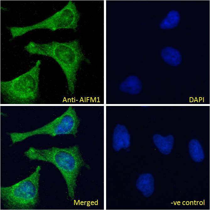
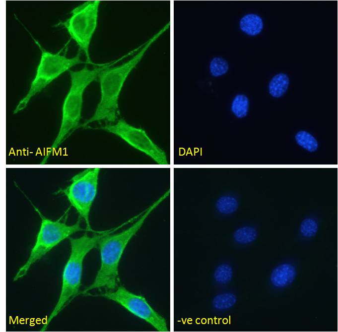
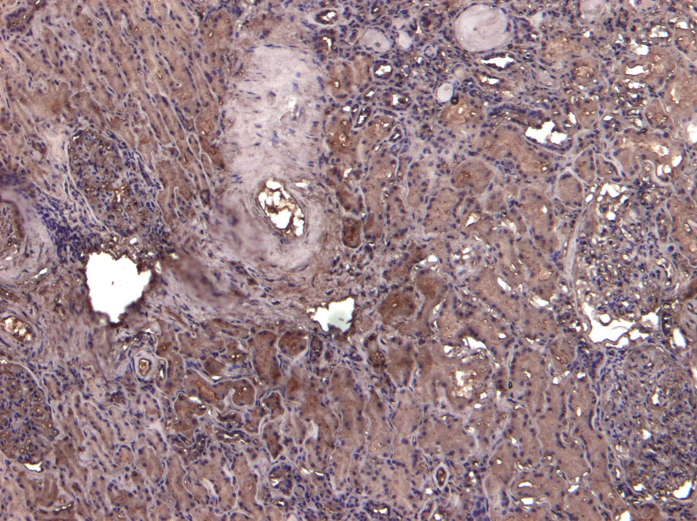
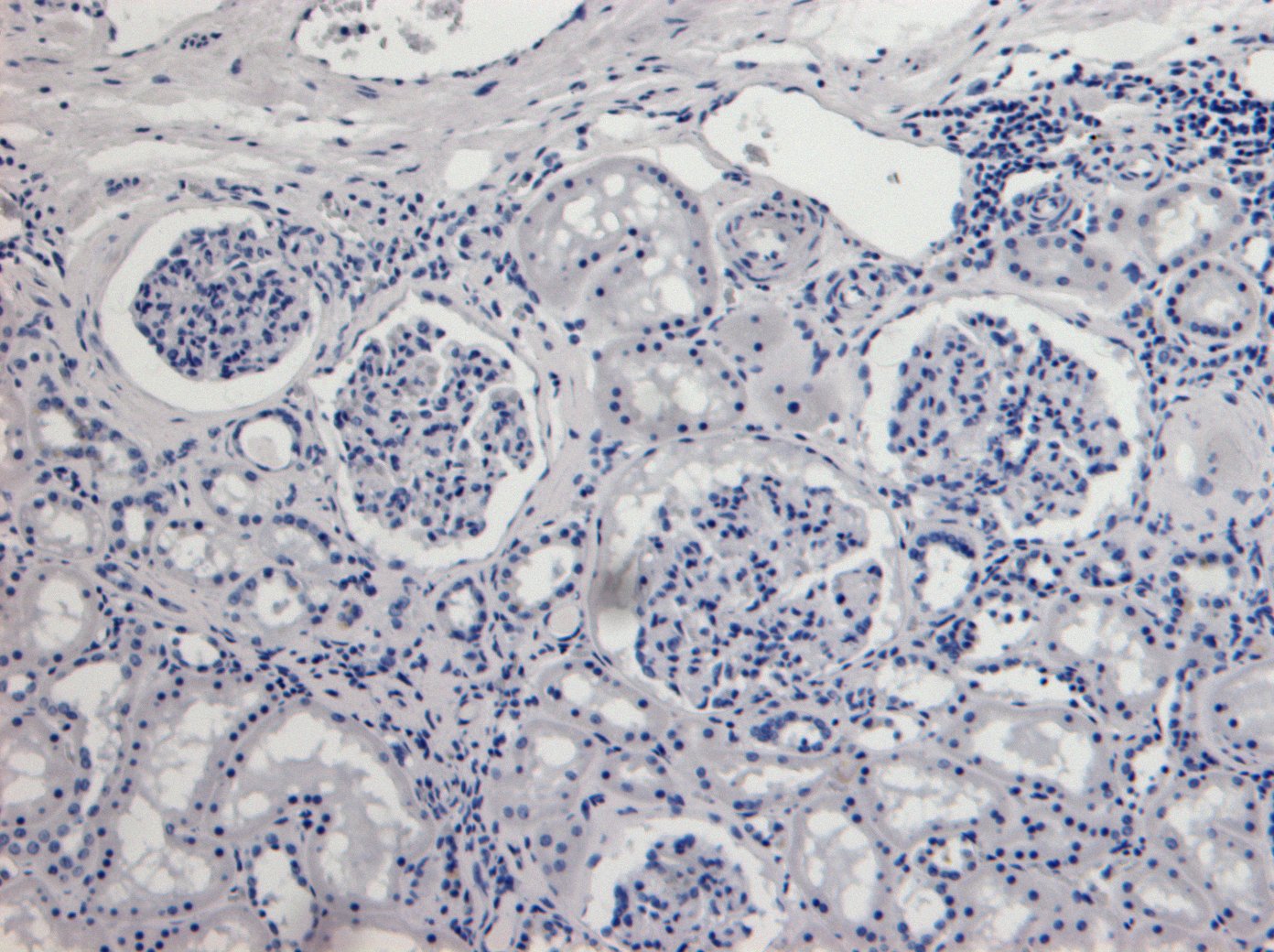
Goat Anti-AIFM1 Antibody (internal region)
Purified Goat Polyclonal Antibody


- SPECIFICATION
- CITATIONS
- PROTOCOLS
- BACKGROUND

Application
| WB, IHC, IF, Pep-ELISA |
|---|---|
| Primary Accession | O95831 |
| Other Accession | NP_004199.1, NP_665811.1, NP_001124319.1 |
| Reactivity | Human |
| Predicted | Human, Cow |
| Host | Goat |
| Clonality | Polyclonal |
| Concentration | 0.5 |
| Calculated MW | 66901 Da |
| Gene ID | 9131 |
|---|---|
| Other Names | AIFM1; apoptosis-inducing factor, mitochondrion-associated, 1; AIF; CMTX4; COWCK; COXPD6; PDCD8; apoptosis-inducing factor 1, mitochondrial; programmed cell death 8 (apoptosis-inducing factor); striatal apoptosis-inducing factor |
| Format | Supplied at 0.5 mg/ml in Tris saline, 0.02% sodium azide, pH7.3 with 0.5% bovine serum albumin. Aliquot and store at -20°C. Minimize freezing and thawing. |
| Immunogen | Peptide with sequence C-DDPNVTKTLRFKQ, from the internal region of the protein sequence according to NP_004199.1; NP_665811.1; NP_001124319.1. |
| Storage | Maintain refrigerated at 2-8°C for up to 6 months. For long term storage store at -20°C in small aliquots to prevent freeze-thaw cycles. |
| Precautions | Goat Anti-AIFM1 Antibody (internal region) is for research use only and not for use in diagnostic or therapeutic procedures. |
| Name | AIFM1 (HGNC:8768) |
|---|---|
| Synonyms | AIF, PDCD8 |
| Function | Functions both as NADH oxidoreductase and as regulator of apoptosis (PubMed:20362274, PubMed:23217327, PubMed:17094969, PubMed:33168626). In response to apoptotic stimuli, it is released from the mitochondrion intermembrane space into the cytosol and to the nucleus, where it functions as a proapoptotic factor in a caspase- independent pathway (PubMed:20362274). Release into the cytoplasm is mediated upon binding to poly-ADP-ribose chains (By similarity). The soluble form (AIFsol) found in the nucleus induces 'parthanatos' i.e. caspase-independent fragmentation of chromosomal DNA (PubMed:20362274). Binds to DNA in a sequence-independent manner (PubMed:27178839). Interacts with EIF3G, and thereby inhibits the EIF3 machinery and protein synthesis, and activates caspase-7 to amplify apoptosis (PubMed:17094969). Plays a critical role in caspase-independent, pyknotic cell death in hydrogen peroxide-exposed cells (PubMed:19418225). In contrast, participates in normal mitochondrial metabolism. Plays an important role in the regulation of respiratory chain biogenesis by interacting with CHCHD4 and controlling CHCHD4 mitochondrial import (PubMed:26004228). |
| Cellular Location | Mitochondrion intermembrane space. Mitochondrion inner membrane. Cytoplasm. Nucleus. Cytoplasm, perinuclear region. Note=Proteolytic cleavage during or just after translocation into the mitochondrial intermembrane space (IMS) results in the formation of an inner-membrane-anchored mature form (AIFmit). During apoptosis, further proteolytic processing leads to a mature form, which is confined to the mitochondrial IMS in a soluble form (AIFsol). AIFsol is released to the cytoplasm in response to specific death signals, and translocated to the nucleus, where it induces nuclear apoptosis (PubMed:15775970). Release into the cytoplasm is mediated upon binding to poly-ADP-ribose chains (By similarity) Translocation into the nucleus is promoted by interaction with (auto- poly-ADP-ribosylated) processed form of PARP1 (PubMed:33168626) Colocalizes with EIF3G in the nucleus and perinuclear region (PubMed:17094969). {ECO:0000250|UniProtKB:Q9Z0X1, ECO:0000269|PubMed:15775970, ECO:0000269|PubMed:17094969, ECO:0000269|PubMed:33168626} [Isoform 4]: Mitochondrion. Cytoplasm, cytosol. Note=In pro-apoptotic conditions, is released from mitochondria to cytosol in a calpain/cathepsin-dependent manner. |
| Tissue Location | Expressed in all tested tissues (PubMed:16644725). Detected in muscle and skin fibroblasts (at protein level) (PubMed:23217327). Expressed in osteoblasts (at protein level) (PubMed:28842795). [Isoform 4]: Expressed in all tested tissues except brain. |

Thousands of laboratories across the world have published research that depended on the performance of antibodies from Abcepta to advance their research. Check out links to articles that cite our products in major peer-reviewed journals, organized by research category.
info@abcepta.com, and receive a free "I Love Antibodies" mug.

Provided below are standard protocols that you may find useful for product applications.
References
The enzymatic activity of apoptosis-inducing factor supports energy metabolism benefiting the growth and invasiveness of advanced prostate cancer cells. Lewis EM, Wilkinson AS, Jackson JS, Mehra R, Varambally S, Chinnaiyan AM, Wilkinson JC. The Journal of biological chemistry 2012 Dec 287 (52): 43862-75.
If you have used an Abcepta product and would like to share how it has performed, please click on the "Submit Review" button and provide the requested information. Our staff will examine and post your review and contact you if needed.
If you have any additional inquiries please email technical services at tech@abcepta.com.
Ordering Information
Other Products
Shipping Information
