


 Foundational characteristics of cancer include proliferation, angiogenesis, migration, evasion of apoptosis, and cellular immortality. Find key markers for these cellular processes and antibodies to detect them.
 The SUMOplot™ Analysis Program predicts and scores sumoylation sites in your protein. SUMOylation is a post-translational modification involved in various cellular processes, such as nuclear-cytosolic transport, transcriptional regulation, apoptosis, protein stability, response to stress, and progression through the cell cycle.
 The Autophagy Receptor Motif Plotter predicts and scores autophagy receptor binding sites in your protein. Identifying proteins connected to this pathway is critical to understanding the role of autophagy in physiological as well as pathological processes such as development, differentiation, neurodegenerative diseases, stress, infection, and cancer.
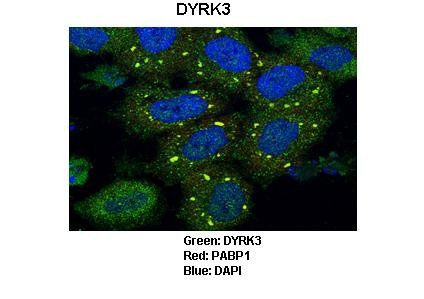
DYRK3 antibody - N-terminal region
Rabbit Polyclonal Antibody

- SPECIFICATION
- CITATIONS
- PROTOCOLS
- BACKGROUND

Application
| WB, IF |
|---|---|
| Primary Accession | O43781 |
| Other Accession | O43781, NP_003573, NM_003582 |
| Reactivity | Human, Mouse |
| Predicted | Human, Mouse |
| Host | Rabbit |
| Clonality | Polyclonal |
| Calculated MW | 66 kDa |
| Gene ID | 8444 |
|---|---|
| Alias Symbol | DYRK5, RED, REDK, hYAK3-2 |
| Other Names | Dual specificity tyrosine-phosphorylation-regulated kinase 3, Regulatory erythroid kinase, REDK, DYRK3 |
| Target/Specificity | This gene product belongs to the DYRK family of dual-specificity protein kinases that catalyze autophosphorylation on serine/threonine and tyrosine residues. The members of this family share structural similarity, however, differ in their substrate specificity, suggesting their involvement in different cellular functions. The encoded protein has been shown to autophosphorylate on tyrosine residue and catalyze phosphorylation of histones H3 and H2B in vitro. Alternatively spliced transcript variants encoding different isoforms have been identified. |
| Format | Liquid. Purified antibody supplied in 1x PBS buffer with 0.09% (w/v) sodium azide and 2% sucrose. |
| Reconstitution & Storage | Add 50 ul of distilled water. Final anti-DYRK3 antibody concentration is 1 mg/ml in PBS buffer with 2% sucrose. For longer periods of storage, store at -20°C. Avoid repeat freeze-thaw cycles. |
| Precautions | DYRK3 antibody - N-terminal region is for research use only and not for use in diagnostic or therapeutic procedures. |
| Name | DYRK3 (HGNC:3094) |
|---|---|
| Function | Dual-specificity protein kinase that promotes disassembly of several types of membraneless organelles during mitosis, such as stress granules, nuclear speckles and pericentriolar material (PubMed:29973724). Dual-specificity tyrosine-regulated kinases (DYRKs) autophosphorylate a critical tyrosine residue in their activation loop and phosphorylate their substrate on serine and threonine residues (PubMed:9748265, PubMed:29634919). Acts as a central dissolvase of membraneless organelles during the G2-to-M transition, after the nuclear-envelope breakdown: acts by mediating phosphorylation of multiple serine and threonine residues in unstructured domains of proteins, such as SRRM1 and PCM1 (PubMed:29973724). Does not mediate disassembly of all membraneless organelles: disassembly of P-body and nucleolus is not regulated by DYRK3 (PubMed:29973724). Dissolution of membraneless organelles at the onset of mitosis is also required to release mitotic regulators, such as ZNF207, from liquid-unmixed organelles where they are sequestered and keep them dissolved during mitosis (PubMed:29973724). Regulates mTORC1 by mediating the dissolution of stress granules: during stressful conditions, DYRK3 partitions from the cytosol to the stress granule, together with mTORC1 components, which prevents mTORC1 signaling (PubMed:23415227). When stress signals are gone, the kinase activity of DYRK3 is required for the dissolution of stress granule and mTORC1 relocation to the cytosol: acts by mediating the phosphorylation of the mTORC1 inhibitor AKT1S1, allowing full reactivation of mTORC1 signaling (PubMed:23415227). Also acts as a negative regulator of EPO-dependent erythropoiesis: may place an upper limit on red cell production during stress erythropoiesis (PubMed:10779429). Inhibits cell death due to cytokine withdrawal in hematopoietic progenitor cells (PubMed:10779429). Promotes cell survival upon genotoxic stress through phosphorylation of SIRT1: this in turn inhibits p53/TP53 activity and apoptosis (PubMed:20167603). |
| Cellular Location | Nucleus. Cytoplasm. Nucleus speckle. Cytoplasmic granule. Cytoplasm, cytoskeleton, microtubule organizing center, centrosome Note=Associates with membraneless organelles in the cytoplasm and nucleus (PubMed:29973724). Shuttles between cytoplasm and stress granules (PubMed:20167603). Localized predominantly on distinct speckles distributed throughout the cytoplasm of the cell (PubMed:20167603). At low concentration, showns a homogeneous distribution throughout the cytoplasm and does not condense in speckles. During oxidative and osmotic stress, localizes to stress granules (PubMed:20167603). |
| Tissue Location | Isoform 1: Highly expressed in testis and in hematopoietic tissue such as fetal liver, and bone marrow (PubMed:10779429). Isoform 1: Predominant form in fetal liver and bone marrow (PubMed:10779429). Isoform 1: Present at low levels in heart, pancreas, lymph node and thymus (PubMed:10779429). Isoform 2: Highly expressed in testis and in hematopoietic tissue such as fetal liver, and bone marrow (PubMed:10779429). Isoform 2: Predominant form in testis. Isoform 2: Present at low levels in heart, pancreas, lymph node and thymus (PubMed:10779429). |

Thousands of laboratories across the world have published research that depended on the performance of antibodies from Abcepta to advance their research. Check out links to articles that cite our products in major peer-reviewed journals, organized by research category.
info@abcepta.com, and receive a free "I Love Antibodies" mug.

Provided below are standard protocols that you may find useful for product applications.
Background
This is a rabbit polyclonal antibody against DYRK3. It was validated on Western Blot using a cell lysate as a positive control. Abgent strives to provide antibodies covering each member of a whole protein family of your interest. We also use our best efforts to provide you antibodies recognize various epitopes of a target protein. For availability of antibody needed for your experiment, please inquire (sales@abgent.com).
If you have used an Abcepta product and would like to share how it has performed, please click on the "Submit Review" button and provide the requested information. Our staff will examine and post your review and contact you if needed.
If you have any additional inquiries please email technical services at tech@abcepta.com.
Ordering Information
Shipping Information
