


 Foundational characteristics of cancer include proliferation, angiogenesis, migration, evasion of apoptosis, and cellular immortality. Find key markers for these cellular processes and antibodies to detect them.
 The SUMOplot™ Analysis Program predicts and scores sumoylation sites in your protein. SUMOylation is a post-translational modification involved in various cellular processes, such as nuclear-cytosolic transport, transcriptional regulation, apoptosis, protein stability, response to stress, and progression through the cell cycle.
 The Autophagy Receptor Motif Plotter predicts and scores autophagy receptor binding sites in your protein. Identifying proteins connected to this pathway is critical to understanding the role of autophagy in physiological as well as pathological processes such as development, differentiation, neurodegenerative diseases, stress, infection, and cancer.
NINJ1 antibody - N-terminal region
Rabbit Polyclonal Antibody
- SPECIFICATION
- CITATIONS
- PROTOCOLS
- BACKGROUND

Application
| WB |
|---|---|
| Primary Accession | Q92982 |
| Other Accession | NM_004148, NP_004139 |
| Reactivity | Human, Mouse, Rat, Horse |
| Predicted | Human, Mouse, Rat |
| Host | Rabbit |
| Clonality | Polyclonal |
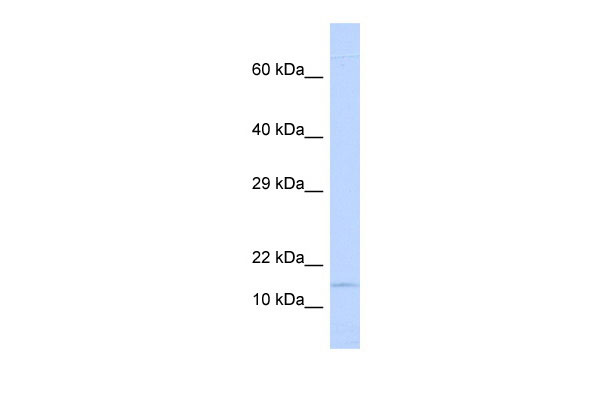
| Calculated MW | 16kDa |
| Gene ID | 4814 |
|---|---|
| Alias Symbol | NIN1, NINJURIN |
| Other Names | Ninjurin-1, Nerve injury-induced protein 1, NINJ1 |
| Format | Liquid. Purified antibody supplied in 1x PBS buffer with 0.09% (w/v) sodium azide and 2% sucrose. |
| Reconstitution & Storage | Add 50 ul of distilled water. Final anti-NINJ1 antibody concentration is 1 mg/ml in PBS buffer with 2% sucrose. For longer periods of storage, store at 20°C. Avoid repeat freeze-thaw cycles. |
| Precautions | NINJ1 antibody - N-terminal region is for research use only and not for use in diagnostic or therapeutic procedures. |
| Name | NINJ1 {ECO:0000303|PubMed:33472215, ECO:0000312|HGNC:HGNC:7824} |
|---|---|
| Function | [Ninjurin-1]: Effector of necroptotic and pyroptotic programmed cell death that mediates plasma membrane rupture (cytolysis) (PubMed:33472215, PubMed:36468682, PubMed:37196676, PubMed:37198476). Acts downstream of Gasdermin (GSDMA, GSDMB, GSDMC, GSDMD, or GSDME) or MLKL during pyroptosis or necroptosis, respectively: oligomerizes in response to death stimuli and promotes plasma membrane rupture by introducing hydrophilic faces of 2 alpha helices into the hydrophobic membrane, leading to release intracellular molecules named damage- associated molecular patterns (DAMPs) that propagate the inflammatory response (PubMed:33472215, PubMed:36468682, PubMed:37196676, PubMed:37198476). Acts as a regulator of Toll-like receptor 4 (TLR4) signaling triggered by lipopolysaccharide (LPS) during systemic inflammation; directly binds LPS (PubMed:26677008). Involved in leukocyte migration during inflammation by promoting transendothelial migration of macrophages via homotypic binding (By similarity). Promotes the migration of monocytes across the brain endothelium to central nervous system inflammatory lesions (PubMed:22162058). Also acts as a homophilic transmembrane adhesion molecule involved in various processes such as axonal growth, cell chemotaxis and angiogenesis (PubMed:8780658, PubMed:9261151, PubMed:33028854). Promotes cell adhesion by mediating homophilic interactions via its extracellular N-terminal adhesion motif (N-NAM) (PubMed:33028854). Involved in the progression of the inflammatory stress by promoting cell-to-cell interactions between immune cells and endothelial cells (PubMed:22162058, PubMed:26677008, PubMed:32147432). Plays a role in nerve regeneration by promoting maturation of Schwann cells (PubMed:8780658, PubMed:9261151). Acts as a regulator of angiogenesis (PubMed:33028854). Promotes the formation of new vessels by mediating the interaction between capillary pericyte cells and endothelial cells (By similarity). Promotes osteoclasts development by enhancing the survival of prefusion osteoclasts (By similarity). Also involved in striated muscle growth and differentiation (By similarity). |
| Cellular Location | [Ninjurin-1]: Cell membrane; Multi-pass membrane protein. Synaptic cell membrane {ECO:0000250|UniProtKB:O70131}; Multi-pass membrane protein |
| Tissue Location | Widely expressed in both adult and embryonic tissues, primarily those of epithelial origin |

Thousands of laboratories across the world have published research that depended on the performance of antibodies from Abcepta to advance their research. Check out links to articles that cite our products in major peer-reviewed journals, organized by research category.
info@abcepta.com, and receive a free "I Love Antibodies" mug.

Provided below are standard protocols that you may find useful for product applications.
References
Guimaraes,P.E.,(er)SpinalCord(2008)InpressReconstitutionandStorage:Forshorttermuse,storeat2-8Cupto1week.Forlongtermstorage,storeat-20Cinsmallaliquotstopreventfreeze-thawcycles.
If you have used an Abcepta product and would like to share how it has performed, please click on the "Submit Review" button and provide the requested information. Our staff will examine and post your review and contact you if needed.
If you have any additional inquiries please email technical services at tech@abcepta.com.
Ordering Information
Other Products
Shipping Information
