


 Foundational characteristics of cancer include proliferation, angiogenesis, migration, evasion of apoptosis, and cellular immortality. Find key markers for these cellular processes and antibodies to detect them.
 The SUMOplot™ Analysis Program predicts and scores sumoylation sites in your protein. SUMOylation is a post-translational modification involved in various cellular processes, such as nuclear-cytosolic transport, transcriptional regulation, apoptosis, protein stability, response to stress, and progression through the cell cycle.
 The Autophagy Receptor Motif Plotter predicts and scores autophagy receptor binding sites in your protein. Identifying proteins connected to this pathway is critical to understanding the role of autophagy in physiological as well as pathological processes such as development, differentiation, neurodegenerative diseases, stress, infection, and cancer.
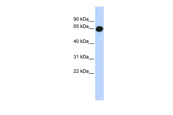
B3GALNT2 antibody - middle region
Rabbit Polyclonal Antibody
- SPECIFICATION
- CITATIONS
- PROTOCOLS
- BACKGROUND

Application
| WB |
|---|---|
| Primary Accession | Q8NCR0 |
| Other Accession | NM_152490, NP_689703 |
| Reactivity | Human, Mouse, Rat, Rabbit, Horse, Bovine, Guinea Pig, Dog |
| Predicted | Human, Rat, Rabbit, Horse, Bovine, Guinea Pig, Dog |
| Host | Rabbit |
| Clonality | Polyclonal |
| Calculated MW | 57kDa |
| Gene ID | 148789 |
|---|---|
| Alias Symbol | B3GalNAc-T2, MGC39558 |
| Other Names | UDP-GalNAc:beta-1, 3-N-acetylgalactosaminyltransferase 2, Beta-1, 3-GalNAc-T2, 2.4.1.-, Beta-1, 3-N-acetylgalactosaminyltransferase II, B3GALNT2 |
| Format | Liquid. Purified antibody supplied in 1x PBS buffer with 0.09% (w/v) sodium azide and 2% sucrose. |
| Reconstitution & Storage | Add 50 ul of distilled water. Final anti-B3GALNT2 antibody concentration is 1 mg/ml in PBS buffer with 2% sucrose. For longer periods of storage, store at 20°C. Avoid repeat freeze-thaw cycles. |
| Precautions | B3GALNT2 antibody - middle region is for research use only and not for use in diagnostic or therapeutic procedures. |
| Name | B3GALNT2 |
|---|---|
| Function | Beta-1,3-N-acetylgalactosaminyltransferase that synthesizes a unique carbohydrate structure, GalNAc-beta-1-3GlcNAc, on N- and O- glycans. Has no galactose nor galactosaminyl transferase activity toward any acceptor substrate. Involved in alpha-dystroglycan (DAG1) glycosylation: acts coordinately with GTDC2/POMGnT2 to synthesize a GalNAc-beta3-GlcNAc-beta-terminus at the 4-position of protein O- mannose in the biosynthesis of the phosphorylated O-mannosyl trisaccharide (N-acetylgalactosamine-beta-3-N-acetylglucosamine-beta-4- (phosphate-6-)mannose), a carbohydrate structure present in alpha- dystroglycan, which is required for binding laminin G-like domain- containing extracellular proteins with high affinity. |
| Cellular Location | Golgi apparatus membrane; Single- pass type II membrane protein. Endoplasmic reticulum |
| Tissue Location | Expressed in all tissues examined, but at highest levels in testis, adipose tissue, skeletal muscle and ovary |

Thousands of laboratories across the world have published research that depended on the performance of antibodies from Abcepta to advance their research. Check out links to articles that cite our products in major peer-reviewed journals, organized by research category.
info@abcepta.com, and receive a free "I Love Antibodies" mug.

Provided below are standard protocols that you may find useful for product applications.
References
Gregory S.G.,et al.Nature 441:315-321(2006).
Totoki Y.,et al.Submitted (MAR-2005) to the EMBL/GenBank/DDBJ databases.
Hiruma T.,et al.J. Biol. Chem. 279:14087-14095(2004).
Stevens E.,et al.Am. J. Hum. Genet. 92:354-365(2013).
Yoshida-Moriguchi T.,et al.Science 341:896-899(2013).
If you have used an Abcepta product and would like to share how it has performed, please click on the "Submit Review" button and provide the requested information. Our staff will examine and post your review and contact you if needed.
If you have any additional inquiries please email technical services at tech@abcepta.com.
Ordering Information
Other Products
Shipping Information
