


 Foundational characteristics of cancer include proliferation, angiogenesis, migration, evasion of apoptosis, and cellular immortality. Find key markers for these cellular processes and antibodies to detect them.
 The SUMOplot™ Analysis Program predicts and scores sumoylation sites in your protein. SUMOylation is a post-translational modification involved in various cellular processes, such as nuclear-cytosolic transport, transcriptional regulation, apoptosis, protein stability, response to stress, and progression through the cell cycle.
 The Autophagy Receptor Motif Plotter predicts and scores autophagy receptor binding sites in your protein. Identifying proteins connected to this pathway is critical to understanding the role of autophagy in physiological as well as pathological processes such as development, differentiation, neurodegenerative diseases, stress, infection, and cancer.
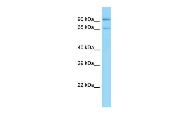
Cdh1 antibody - C-terminal region
Rabbit Polyclonal Antibody
- SPECIFICATION
- CITATIONS
- PROTOCOLS
- BACKGROUND

Application
| WB |
|---|---|
| Primary Accession | P09803 |
| Other Accession | NM_009864, NP_033994 |
| Reactivity | Human, Mouse, Rat, Rabbit, Pig, Horse, Bovine, Guinea Pig, Dog |
| Predicted | Human, Mouse, Rabbit, Pig, Chicken, Horse, Bovine, Guinea Pig, Dog |
| Host | Rabbit |
| Clonality | Polyclonal |
| Calculated MW | 98kDa |
| Gene ID | 12550 |
|---|---|
| Alias Symbol | AA960649, Ecad, L-CAM, MGC107495, UVO, Um |
| Other Names | Cadherin-1, ARC-1, Epithelial cadherin, E-cadherin, Uvomorulin, CD324, E-Cad/CTF1, E-Cad/CTF2, E-Cad/CTF3, Cdh1 |
| Format | Liquid. Purified antibody supplied in 1x PBS buffer with 0.09% (w/v) sodium azide and 2% sucrose. |
| Reconstitution & Storage | Add 50 ul of distilled water. Final anti-Cdh1 antibody concentration is 1 mg/ml in PBS buffer with 2% sucrose. For longer periods of storage, store at 20°C. Avoid repeat freeze-thaw cycles. |
| Precautions | Cdh1 antibody - C-terminal region is for research use only and not for use in diagnostic or therapeutic procedures. |
| Name | Cdh1 |
|---|---|
| Function | Cadherins are calcium-dependent cell adhesion proteins (PubMed:11976333). They preferentially interact with themselves in a homophilic manner in connecting cells; cadherins may thus contribute to the sorting of heterogeneous cell types. CDH1 is involved in mechanisms regulating cell-cell adhesions, mobility and proliferation of epithelial cells (PubMed:11976333). Has a potent invasive suppressor role. It is a ligand for integrin alpha-E/beta-7 (By similarity). |
| Cellular Location | Cell junction, adherens junction. Cell membrane; Single-pass type I membrane protein. Endosome. Golgi apparatus, trans-Golgi network. Note=Colocalizes with DLGAP5 at sites of cell- cell contact in intestinal epithelial cells. Anchored to actin microfilaments through association with alpha-, beta- and gamma- catenin. Sequential proteolysis induced by apoptosis or calcium influx, results in translocation from sites of cell-cell contact to the cytoplasm. Colocalizes with RAB11A endosomes during its transport from the Golgi apparatus to the plasma membrane (By similarity) |
| Tissue Location | Expressed in inner and outer pillar cells of the organ of Corti (at protein level) (PubMed:30639848). Non-neural epithelial tissues. |

Thousands of laboratories across the world have published research that depended on the performance of antibodies from Abcepta to advance their research. Check out links to articles that cite our products in major peer-reviewed journals, organized by research category.
info@abcepta.com, and receive a free "I Love Antibodies" mug.

Provided below are standard protocols that you may find useful for product applications.
References
Nagafuchi A.,et al.Nature 329:341-343(1987).
Ringwald M.,et al.Nucleic Acids Res. 19:6533-6539(1991).
Ringwald M.,et al.EMBO J. 6:3647-3653(1987).
Behrens J.,et al.Proc. Natl. Acad. Sci. U.S.A. 88:11495-11499(1991).
Butz S.,et al.FEBS Lett. 355:195-200(1994).
If you have used an Abcepta product and would like to share how it has performed, please click on the "Submit Review" button and provide the requested information. Our staff will examine and post your review and contact you if needed.
If you have any additional inquiries please email technical services at tech@abcepta.com.
Ordering Information
Other Products
Shipping Information
