


 Foundational characteristics of cancer include proliferation, angiogenesis, migration, evasion of apoptosis, and cellular immortality. Find key markers for these cellular processes and antibodies to detect them.
 The SUMOplot™ Analysis Program predicts and scores sumoylation sites in your protein. SUMOylation is a post-translational modification involved in various cellular processes, such as nuclear-cytosolic transport, transcriptional regulation, apoptosis, protein stability, response to stress, and progression through the cell cycle.
 The Autophagy Receptor Motif Plotter predicts and scores autophagy receptor binding sites in your protein. Identifying proteins connected to this pathway is critical to understanding the role of autophagy in physiological as well as pathological processes such as development, differentiation, neurodegenerative diseases, stress, infection, and cancer.
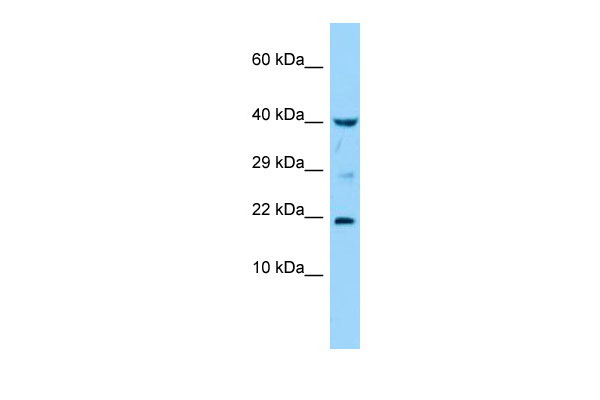
IL36G Antibody - middle region
Rabbit Polyclonal Antibody
- SPECIFICATION
- CITATIONS
- PROTOCOLS
- BACKGROUND

Application
| WB |
|---|---|
| Primary Accession | Q9NZH8 |
| Other Accession | NM_019618, NP_062564 |
| Reactivity | Human, Rabbit, Horse |
| Predicted | Human, Rabbit, Horse |
| Host | Rabbit |
| Clonality | Polyclonal |
| Calculated MW | 19kDa |
| Gene ID | 56300 |
|---|---|
| Alias Symbol | IL-1F9, IL-1H1, IL-1RP2, IL1E, IL1F9, IL1H1, IL1RP2 |
| Other Names | Interleukin-36 gamma, IL-1-related protein 2, IL-1RP2, Interleukin-1 epsilon, IL-1 epsilon, Interleukin-1 family member 9, IL-1F9, Interleukin-1 homolog 1, IL-1H1, IL36G, IL1E, IL1F9, IL1H1, IL1RP2 |
| Format | Liquid. Purified antibody supplied in 1x PBS buffer with 0.09% (w/v) sodium azide and 2% sucrose. |
| Reconstitution & Storage | Add 50 ul of distilled water. Final anti-IL36G antibody concentration is 1 mg/ml in PBS buffer with 2% sucrose. For longer periods of storage, store at 20°C. Avoid repeat freeze-thaw cycles. |
| Precautions | IL36G Antibody - middle region is for research use only and not for use in diagnostic or therapeutic procedures. |
| Name | IL36G (HGNC:15741) |
|---|---|
| Function | Cytokine that binds to and signals through the IL1RL2/IL-36R receptor which in turn activates NF-kappa-B and MAPK signaling pathways in target cells. Part of the IL-36 signaling system that is thought to be present in epithelial barriers and to take part in local inflammatory response; similar to the IL-1 system with which it shares the coreceptor IL1RAP. Seems to be involved in skin inflammatory response by acting on keratinocytes, dendritic cells and indirectly on T-cells to drive tissue infiltration, cell maturation and cell proliferation. In cultured keratinocytes induces the expression of macrophage, T-cell, and neutrophil chemokines, such as CCL3, CCL4, CCL5, CCL2, CCL17, CCL22, CL20, CCL5, CCL2, CCL17, CCL22, CXCL8, CCL20 and CXCL1; also stimulates its own expression and that of the prototypic cutaneous pro-inflammatory parameters TNF-alpha, S100A7/psoriasin and inducible NOS. May play a role in pro-inflammatory responses during particular neutrophilic airway inflammation: activates mitogen-activated protein kinases and NF-kappa B in primary lung fibroblasts, and stimulates the expression of IL-8 and CXCL3 and Th17 chemokine CCL20 in lung fibroblasts. May be involved in the innate immune response to fungal pathogens, such as Aspergillus fumigatus. |
| Cellular Location | Cytoplasm. Secreted. Note=The secretion is dependent on protein unfolding and facilitated by the cargo receptor TMED10; it results in protein translocation from the cytoplasm into the ERGIC (endoplasmic reticulum-Golgi intermediate compartment) followed by vesicle entry and secretion. |
| Tissue Location | Highly expressed in tissues containing epithelial cells: skin, lung, stomach and esophagus. Expressed in bronchial epithelial. In skin is expressed only in keratinocytes but not in fibroblasts, endothelial cells or melanocytes. Up-regulated in lesional psoriasis skin. Expressed in monocyte-derived dendritic cells and M1 macrophages. |

Thousands of laboratories across the world have published research that depended on the performance of antibodies from Abcepta to advance their research. Check out links to articles that cite our products in major peer-reviewed journals, organized by research category.
info@abcepta.com, and receive a free "I Love Antibodies" mug.

Provided below are standard protocols that you may find useful for product applications.
References
Kumar S.,et al.J. Biol. Chem. 275:10308-10314(2000).
Debets R.,et al.J. Immunol. 167:1440-1446(2001).
Busfield S.J.,et al.Genomics 66:213-216(2000).
Nicklin M.J.H.,et al.Genomics 79:718-725(2002).
Clark H.F.,et al.Genome Res. 13:2265-2270(2003).
If you have used an Abcepta product and would like to share how it has performed, please click on the "Submit Review" button and provide the requested information. Our staff will examine and post your review and contact you if needed.
If you have any additional inquiries please email technical services at tech@abcepta.com.
Ordering Information
Other Products
Shipping Information
