


 Foundational characteristics of cancer include proliferation, angiogenesis, migration, evasion of apoptosis, and cellular immortality. Find key markers for these cellular processes and antibodies to detect them.
 The SUMOplot™ Analysis Program predicts and scores sumoylation sites in your protein. SUMOylation is a post-translational modification involved in various cellular processes, such as nuclear-cytosolic transport, transcriptional regulation, apoptosis, protein stability, response to stress, and progression through the cell cycle.
 The Autophagy Receptor Motif Plotter predicts and scores autophagy receptor binding sites in your protein. Identifying proteins connected to this pathway is critical to understanding the role of autophagy in physiological as well as pathological processes such as development, differentiation, neurodegenerative diseases, stress, infection, and cancer.
MMP13 Antibody (C-Terminus)
Rabbit Polyclonal Antibody
- SPECIFICATION
- CITATIONS: 1
- PROTOCOLS
- BACKGROUND

Application
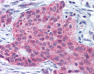
| IHC-P |
|---|---|
| Primary Accession | P45452 |
| Reactivity | Human |
| Host | Rabbit |
| Clonality | Polyclonal |
| Calculated MW | 54kDa |
| Dilution | IHC-P (5 µg/ml) |
| Gene ID | 4322 |
|---|---|
| Other Names | Collagenase 3, 3.4.24.-, Matrix metalloproteinase-13, MMP-13, MMP13 |
| Target/Specificity | Human MMP13. BLAST analysis of the peptide immunogen showed no homology with other human proteins. |
| Reconstitution & Storage | Store at 4°C for short term applications. For long term storage, aliquot and store at -20°C. |
| Precautions | MMP13 Antibody (C-Terminus) is for research use only and not for use in diagnostic or therapeutic procedures. |
| Name | MMP13 |
|---|---|
| Function | Plays a role in the degradation of extracellular matrix proteins including fibrillar collagen, fibronectin, TNC and ACAN. Cleaves triple helical collagens, including type I, type II and type III collagen, but has the highest activity with soluble type II collagen. Can also degrade collagen type IV, type XIV and type X. May also function by activating or degrading key regulatory proteins, such as TGFB1 and CCN2. Plays a role in wound healing, tissue remodeling, cartilage degradation, bone development, bone mineralization and ossification. Required for normal embryonic bone development and ossification. Plays a role in the healing of bone fractures via endochondral ossification. Plays a role in wound healing, probably by a mechanism that involves proteolytic activation of TGFB1 and degradation of CCN2. Plays a role in keratinocyte migration during wound healing. May play a role in cell migration and in tumor cell invasion. |
| Cellular Location | Secreted, extracellular space, extracellular matrix. Secreted |
| Tissue Location | Detected in fetal cartilage and calvaria, in chondrocytes of hypertrophic cartilage in vertebrae and in the dorsal end of ribs undergoing ossification, as well as in osteoblasts and periosteal cells below the inner periosteal region of ossified ribs Detected in chondrocytes from in joint cartilage that have been treated with TNF and IL1B, but not in untreated chondrocytes. Detected in T lymphocytes. Detected in breast carcinoma tissue |
| Volume | 50 µl |

Provided below are standard protocols that you may find useful for product applications.
Background
Plays a role in the degradation of extracellular matrix proteins including fibrillar collagen, fibronectin, TNC and ACAN. Cleaves triple helical collagens, including type I, type II and type III collagen, but has the highest activity with soluble type II collagen. Can also degrade collagen type IV, type XIV and type X. May also function by activating or degrading key regulatory proteins, such as TGFB1 and CTGF. Plays a role in wound healing, tissue remodeling, cartilage degradation, bone development, bone mineralization and ossification. Required for normal embryonic bone development and ossification. Plays a role in the healing of bone fractures via endochondral ossification. Plays a role in wound healing, probably by a mechanism that involves proteolytic activation of TGFB1 and degradation of CTGF. Plays a role in keratinocyte migration during wound healing. May play a role in cell migration and in tumor cell invasion.
References
Freije J.M.P.,et al.J. Biol. Chem. 269:16766-16773(1994).
Willmroth F.,et al.Immunobiology 198:375-384(1998).
Ota T.,et al.Nat. Genet. 36:40-45(2004).
Knaeuper V.,et al.J. Biol. Chem. 271:1544-1550(1996).
Knaeuper V.,et al.J. Biol. Chem. 271:17124-17131(1996).
If you have used an Abcepta product and would like to share how it has performed, please click on the "Submit Review" button and provide the requested information. Our staff will examine and post your review and contact you if needed.
If you have any additional inquiries please email technical services at tech@abcepta.com.
Ordering Information
Other Products
Shipping Information
