


 Foundational characteristics of cancer include proliferation, angiogenesis, migration, evasion of apoptosis, and cellular immortality. Find key markers for these cellular processes and antibodies to detect them.
 The SUMOplot™ Analysis Program predicts and scores sumoylation sites in your protein. SUMOylation is a post-translational modification involved in various cellular processes, such as nuclear-cytosolic transport, transcriptional regulation, apoptosis, protein stability, response to stress, and progression through the cell cycle.
 The Autophagy Receptor Motif Plotter predicts and scores autophagy receptor binding sites in your protein. Identifying proteins connected to this pathway is critical to understanding the role of autophagy in physiological as well as pathological processes such as development, differentiation, neurodegenerative diseases, stress, infection, and cancer.
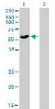
MYOC / Myocilin Antibody (clone 4F8)
Mouse Monoclonal Antibody
- SPECIFICATION
- CITATIONS
- PROTOCOLS
- BACKGROUND

Application
| WB, IHC-P, E, RNAi |
|---|---|
| Primary Accession | Q99972 |
| Reactivity | Human |
| Host | Mouse |
| Clonality | Monoclonal |
| Clone Names | 4F8 |
| Calculated MW | 57kDa |
| Dilution | IHC-P (5 µg/ml) |
| Gene ID | 4653 |
|---|---|
| Other Names | Myocilin, Myocilin 55 kDa subunit, Trabecular meshwork-induced glucocorticoid response protein, Myocilin, N-terminal fragment, Myocilin 20 kDa N-terminal fragment, Myocilin, C-terminal fragment, Myocilin 35 kDa N-terminal fragment, MYOC, GLC1A, TIGR |
| Reconstitution & Storage | Store at -20°C. Aliquot to avoid freeze/thaw cycles. |
| Precautions | MYOC / Myocilin Antibody (clone 4F8) is for research use only and not for use in diagnostic or therapeutic procedures. |
| Name | MYOC |
|---|---|
| Synonyms | GLC1A, TIGR {ECO:0000303|PubMed:9280311} |
| Function | Secreted glycoprotein regulating the activation of different signaling pathways in adjacent cells to control different processes including cell adhesion, cell-matrix adhesion, cytoskeleton organization and cell migration. Promotes substrate adhesion, spreading and formation of focal contacts. Negatively regulates cell-matrix adhesion and stress fiber assembly through Rho protein signal transduction. Modulates the organization of actin cytoskeleton by stimulating the formation of stress fibers through interactions with components of Wnt signaling pathways. Promotes cell migration through activation of PTK2 and the downstream phosphatidylinositol 3-kinase signaling. Plays a role in bone formation and promotes osteoblast differentiation in a dose-dependent manner through mitogen-activated protein kinase signaling. Mediates myelination in the peripheral nervous system through ERBB2/ERBB3 signaling. Plays a role as a regulator of muscle hypertrophy through the components of dystrophin- associated protein complex. Involved in positive regulation of mitochondrial depolarization. Plays a role in neurite outgrowth. May participate in the obstruction of fluid outflow in the trabecular meshwork. |
| Cellular Location | Secreted. Golgi apparatus. Cytoplasmic vesicle. Secreted, extracellular space. Secreted, extracellular space, extracellular matrix. Secreted, extracellular exosome. Mitochondrion. Mitochondrion intermembrane space. Mitochondrion inner membrane. Mitochondrion outer membrane. Rough endoplasmic reticulum. Cell projection. Cell projection, cilium. Note=Located preferentially in the ciliary rootlet and basal body of the connecting cilium of photoreceptor cells, and in the rough endoplasmic reticulum (PubMed:9169133). It is only imported to mitochondria in the trabecular meshwork (PubMed:17516541). Localizes to the Golgi apparatus in Schlemm's canal endothelial cells (PubMed:11053284). Appears in the extracellular space of trabecular meshwork cells by an unconventional mechanism, likely associated with exosome-like vesicles (PubMed:15944158). Localizes in trabecular meshwork extracellular matrix (PubMed:15944158). [Myocilin, N-terminal fragment]: Endoplasmic reticulum. Note=Remains retained in the endoplasmic reticulum |
| Tissue Location | Detected in aqueous humor (PubMed:12697062). Detected in the eye (at protein level) (PubMed:11431441). Widely expressed. Highly expressed in various types of muscle, ciliary body, papillary sphincter, skeletal muscle, heart, and bone marrow-derived mesenchymal stem cells. Expressed predominantly in the retina. In normal eyes, found in the inner uveal meshwork region and the anterior portion of the meshwork. In contrast, in many glaucomatous eyes, it is found in more regions of the meshwork and seems to be expressed at higher levels than in normal eyes, regardless of the type or clinical severity of glaucoma. The myocilin 35 kDa fragment is detected in aqueous humor and to a lesser extent in iris and ciliary body |

Thousands of laboratories across the world have published research that depended on the performance of antibodies from Abcepta to advance their research. Check out links to articles that cite our products in major peer-reviewed journals, organized by research category.
info@abcepta.com, and receive a free "I Love Antibodies" mug.

Provided below are standard protocols that you may find useful for product applications.
Background
Secreted glycoprotein regulating the activation of different signaling pathways in adjacent cells to control different processes including cell adhesion, cell-matrix adhesion, cytoskeleton organization and cell migration. Promotes substrate adhesion, spreading and formation of focal contacts. Negatively regulates cell-matrix adhesion and stress fiber assembly through Rho protein signal transduction. Modulates the organization of actin cytoskeleton by stimulating the formation of stress fibers through interactions with components of Wnt signaling pathways. Promotes cell migration through activation of PTK2 and the downstream phosphatidylinositol 3-kinase signaling. Plays a role in bone formation and promotes osteoblast differentiation in a dose-dependent manner through mitogen-activated protein kinase signaling. Mediates myelination in the peripheral nervous system through ERBB2/ERBB3 signaling. Plays a role as a regulator of muscle hypertrophy through the components of dystrophin-associated protein complex. Involved in positive regulation of mitochondrial depolarization. Plays a role in neurite outgrowth. May participate in the obstruction of fluid outflow in the trabecular meshwork.
References
Ortego J.,et al.FEBS Lett. 413:349-353(1997).
Kubota R.,et al.Genomics 41:360-369(1997).
Adam M.F.,et al.Hum. Mol. Genet. 6:2091-2097(1997).
Stone E.M.,et al.Science 275:668-670(1997).
Kubota R.,et al.Biochem. Biophys. Res. Commun. 242:396-400(1998).
If you have used an Abcepta product and would like to share how it has performed, please click on the "Submit Review" button and provide the requested information. Our staff will examine and post your review and contact you if needed.
If you have any additional inquiries please email technical services at tech@abcepta.com.
Ordering Information
Other Products
Shipping Information
