


 Foundational characteristics of cancer include proliferation, angiogenesis, migration, evasion of apoptosis, and cellular immortality. Find key markers for these cellular processes and antibodies to detect them.
 The SUMOplot™ Analysis Program predicts and scores sumoylation sites in your protein. SUMOylation is a post-translational modification involved in various cellular processes, such as nuclear-cytosolic transport, transcriptional regulation, apoptosis, protein stability, response to stress, and progression through the cell cycle.
 The Autophagy Receptor Motif Plotter predicts and scores autophagy receptor binding sites in your protein. Identifying proteins connected to this pathway is critical to understanding the role of autophagy in physiological as well as pathological processes such as development, differentiation, neurodegenerative diseases, stress, infection, and cancer.
Syndapin I / PACSIN1 Antibody
Goat Polyclonal Antibody

- SPECIFICATION
- CITATIONS
- PROTOCOLS
- BACKGROUND

Application
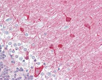
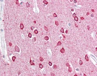
| WB, IHC-P, E |
|---|---|
| Primary Accession | Q9BY11 |
| Reactivity | Human, Pig |
| Host | Goat |
| Clonality | Polyclonal |
| Calculated MW | 51kDa |
| Dilution | ELISA (1:32000), IHC-P (2.5-3.75 µg/ml), WB (0.03-0.1 µg/ml) |
| Gene ID | 29993 |
|---|---|
| Other Names | Protein kinase C and casein kinase substrate in neurons protein 1, Syndapin-1, PACSIN1, KIAA1379 |
| Target/Specificity | Human PACSIN1. Reported variants represent identical protein (NP_065855.1; NP_001186512.1). |
| Reconstitution & Storage | Store at -20°C. Minimize freezing and thawing. |
| Precautions | Syndapin I / PACSIN1 Antibody is for research use only and not for use in diagnostic or therapeutic procedures. |
| Name | PACSIN1 |
|---|---|
| Synonyms | KIAA1379 |
| Function | Plays a role in the reorganization of the microtubule cytoskeleton via its interaction with MAPT; this decreases microtubule stability and inhibits MAPT-induced microtubule polymerization. Plays a role in cellular transport processes by recruiting DNM1, DNM2 and DNM3 to membranes. Plays a role in the reorganization of the actin cytoskeleton and in neuron morphogenesis via its interaction with COBL and WASL, and by recruiting COBL to the cell cortex. Plays a role in the regulation of neurite formation, neurite branching and the regulation of neurite length. Required for normal synaptic vesicle endocytosis; this process retrieves previously released neurotransmitters to accommodate multiple cycles of neurotransmission. Required for normal excitatory and inhibitory synaptic transmission (By similarity). Binds to membranes via its F-BAR domain and mediates membrane tubulation. |
| Cellular Location | Cytoplasm. Cell projection. Synapse, synaptosome. Cell projection, ruffle membrane. Membrane; Peripheral membrane protein Cytoplasmic vesicle membrane; Peripheral membrane protein. Synapse. Cytoplasm, cytosol Cell membrane; Peripheral membrane protein; Cytoplasmic side. Note=Colocalizes with MAPT in axons. In primary neuronal cultures, present at a high level in presynaptic nerve terminals and in the cell body. Colocalizes with DNM1 at vesicular structures in the cell body and neurites (By similarity). Associates with membranes via its F-BAR domain. |
| Tissue Location | Highly expressed in brain and, at much lower levels, in heart and pancreas. |

Thousands of laboratories across the world have published research that depended on the performance of antibodies from Abcepta to advance their research. Check out links to articles that cite our products in major peer-reviewed journals, organized by research category.
info@abcepta.com, and receive a free "I Love Antibodies" mug.

Provided below are standard protocols that you may find useful for product applications.
Background
Plays a role in the reorganization of the microtubule cytoskeleton via its interaction with MAPT; this decreases microtubule stability and inhibits MAPT-induced microtubule polymerization. Plays a role in cellular transport processes by recruiting DNM1, DNM2 and DNM3 to membranes. Plays a role in the reorganization of the actin cytoskeleton and in neuron morphogenesis via its interaction with COBL and WASL, and by recruiting COBL to the cell cortex. Plays a role in the regulation of neurite formation, neurite branching and the regulation of neurite length. Required for normal synaptic vesicle endocytosis; this process retrieves previously released neurotransmitters to accommodate multiple cycles of neurotransmission. Required for normal excitatory and inhibitory synaptic transmission (By similarity). Binds to membranes via its F-BAR domain and mediates membrane tubulation.
References
Sumoy L.,et al.Gene 262:199-205(2001).
Nagase T.,et al.DNA Res. 7:65-73(2000).
Nakajima D.,et al.DNA Res. 9:99-106(2002).
Bechtel S.,et al.BMC Genomics 8:399-399(2007).
Goh S.L.,et al.PLoS ONE 7:E51628-E51628(2012).
If you have used an Abcepta product and would like to share how it has performed, please click on the "Submit Review" button and provide the requested information. Our staff will examine and post your review and contact you if needed.
If you have any additional inquiries please email technical services at tech@abcepta.com.
Ordering Information
Other Products
Shipping Information
