


 Foundational characteristics of cancer include proliferation, angiogenesis, migration, evasion of apoptosis, and cellular immortality. Find key markers for these cellular processes and antibodies to detect them.
 The SUMOplot™ Analysis Program predicts and scores sumoylation sites in your protein. SUMOylation is a post-translational modification involved in various cellular processes, such as nuclear-cytosolic transport, transcriptional regulation, apoptosis, protein stability, response to stress, and progression through the cell cycle.
 The Autophagy Receptor Motif Plotter predicts and scores autophagy receptor binding sites in your protein. Identifying proteins connected to this pathway is critical to understanding the role of autophagy in physiological as well as pathological processes such as development, differentiation, neurodegenerative diseases, stress, infection, and cancer.
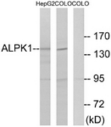
ALPK1 Antibody (aa11-60)
Rabbit Polyclonal Antibody
- SPECIFICATION
- CITATIONS
- PROTOCOLS
- BACKGROUND

Application
| WB, IHC-P, E |
|---|---|
| Primary Accession | Q96QP1 |
| Other Accession | 80216 |
| Reactivity | Human, Mouse |
| Host | Rabbit |
| Clonality | Polyclonal |
| Isotype | IgG |
| Calculated MW | 138861 Da |
| Dilution | ELISA (1:20000), IHC-P (10 µg/ml), WB (1:500 - 1:1000), |
| Gene ID | 80216 |
|---|---|
| Other Names | ALPK1, Alpha-protein kinase 1, Chromosome 4 kinase, LAK, Lymphocyte alpha-kinase, KIAA1527, Alpha-kinase 1 |
| Target/Specificity | ALPK1 Antibody detects endogenous levels of total ALPK1 protein. |
| Reconstitution & Storage | PBS (without Mg2+, Ca2+), pH 7.4, 150 mM sodium chloride, 0.02% sodium azide, 50% glycerol. Store at -20°C for up to one year. |
| Precautions | ALPK1 Antibody (aa11-60) is for research use only and not for use in diagnostic or therapeutic procedures. |
| Name | ALPK1 {ECO:0000303|PubMed:30111836, ECO:0000312|HGNC:HGNC:20917} |
|---|---|
| Function | Serine/threonine-protein kinase that detects bacterial pathogen-associated molecular pattern metabolites (PAMPs) and initiates an innate immune response, a critical step for pathogen elimination and engagement of adaptive immunity (PubMed:28877472, PubMed:28222186, PubMed:30111836). Specifically recognizes and binds ADP-D-glycero-beta- D-manno-heptose (ADP-Heptose), a potent PAMP present in all Gram- negative and some Gram-positive bacteria (PubMed:30111836). ADP- Heptose-binding stimulates its kinase activity to phosphorylate and activate TIFA, triggering pro-inflammatory NF-kappa-B signaling (PubMed:30111836). May be involved in monosodium urate monohydrate (MSU)-induced inflammation by mediating phosphorylation of unconventional myosin MYO9A (PubMed:27169898). May also play a role in apical protein transport by mediating phosphorylation of unconventional myosin MYO1A (PubMed:15883161). May play a role in ciliogenesis (PubMed:30967659). |
| Cellular Location | Cytoplasm, cytosol. Cytoplasm, cytoskeleton, spindle pole Cytoplasm, cytoskeleton, microtubule organizing center, centrosome. Cell projection, cilium. Note=Localized at the base of primary cilia. |
| Tissue Location | Highly expressed in liver. Expressed in the optic nerve and retinal pigmented epithelium. Lower expression is observed in the macula and extramacular retina (PubMed:30967659) |
| Volume | 50 µl |

Thousands of laboratories across the world have published research that depended on the performance of antibodies from Abcepta to advance their research. Check out links to articles that cite our products in major peer-reviewed journals, organized by research category.
info@abcepta.com, and receive a free "I Love Antibodies" mug.

Provided below are standard protocols that you may find useful for product applications.
Background
Kinase that recognizes phosphorylation sites in which the surrounding peptides have an alpha-helical conformation.
References
Ryazanov A.G.,et al.Curr. Biol. 9:R43-R45(1999).
Ota T.,et al.Nat. Genet. 36:40-45(2004).
Hillier L.W.,et al.Nature 434:724-731(2005).
Jikuya H.,et al.Submitted (SEP-2003) to the EMBL/GenBank/DDBJ databases.
Nagase T.,et al.DNA Res. 7:143-150(2000).
If you have used an Abcepta product and would like to share how it has performed, please click on the "Submit Review" button and provide the requested information. Our staff will examine and post your review and contact you if needed.
If you have any additional inquiries please email technical services at tech@abcepta.com.
Ordering Information
Shipping Information
