


 Foundational characteristics of cancer include proliferation, angiogenesis, migration, evasion of apoptosis, and cellular immortality. Find key markers for these cellular processes and antibodies to detect them.
 The SUMOplot™ Analysis Program predicts and scores sumoylation sites in your protein. SUMOylation is a post-translational modification involved in various cellular processes, such as nuclear-cytosolic transport, transcriptional regulation, apoptosis, protein stability, response to stress, and progression through the cell cycle.
 The Autophagy Receptor Motif Plotter predicts and scores autophagy receptor binding sites in your protein. Identifying proteins connected to this pathway is critical to understanding the role of autophagy in physiological as well as pathological processes such as development, differentiation, neurodegenerative diseases, stress, infection, and cancer.
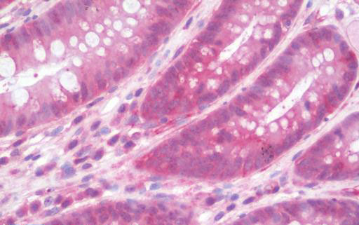
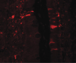
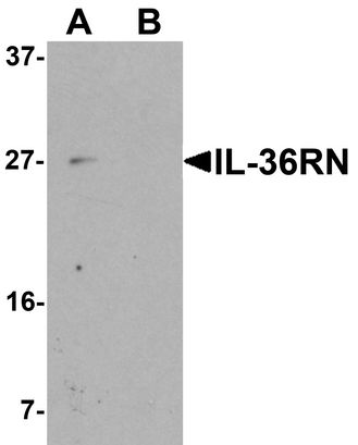
IL36RN / IL1F5 Antibody (C-Terminus)
Rabbit Polyclonal Antibody
- SPECIFICATION
- CITATIONS
- PROTOCOLS
- BACKGROUND

Application
| WB, IHC-P, IF, E |
|---|---|
| Primary Accession | Q9UBH0 |
| Other Accession | 26525 |
| Reactivity | Human, Mouse |
| Host | Rabbit |
| Clonality | Polyclonal |
| Isotype | IgG |
| Calculated MW | 16962 Da |
| Dilution | IF (20 µg/ml), IHC-P (10 µg/ml), WB (1 - 2 µg/ml), |
| Gene ID | 26525 |
|---|---|
| Other Names | IL36RN, FIL1, FIL1D, IL-1 delta, IL-1 related protein 3, IL-1HY1, IL-1RP3, IL-1-related protein 3, IL1F5, IL1RP3, IL1L1, IL36RA, Interleukin-1 delta, Family of interleukin 1-delta, FIL1 delta, IL-1L1, IL-1ra homolog 1, Interleukin-1 family member 5, ... |
| Target/Specificity | IL-36RN antibody is human specific. At least two isoforms of IL-36RN are known to exist. |
| Reconstitution & Storage | PBS, 0.02% sodium azide. Long term: -20°C; Short term: +4°C. Avoid repeat freeze-thaw cycles. |
| Precautions | IL36RN / IL1F5 Antibody (C-Terminus) is for research use only and not for use in diagnostic or therapeutic procedures. |
| Name | IL36RN (HGNC:15561) |
|---|---|
| Function | Inhibits the activity of interleukin-36 (IL36A,IL36B and IL36G) by binding to receptor IL1RL2 and preventing its association with the coreceptor IL1RAP for signaling. Part of the IL-36 signaling system that is thought to be present in epithelial barriers and to take part in local inflammatory response; similar to the IL-1 system with which it shares the coreceptor. Proposed to play a role in skin inflammation. May be involved in the innate immune response to fungal pathogens, such as Aspergillus fumigatus. May activate an anti- inflammatory signaling pathway by recruiting SIGIRR. |
| Cellular Location | Cytoplasm. Secreted. Note=The secretion is dependent on protein unfolding and facilitated by the cargo receptor TMED10; it results in protein translocation from the cytoplasm into the ERGIC (endoplasmic reticulum-Golgi intermediate compartment) followed by vesicle entry and secretion. |
| Tissue Location | Predominantly expressed in skin keratinocytes but not in fibroblasts, endothelial cells or melanocytes. Detected also in the spleen, brain leukocyte and macrophage cell types. Increased in lesional psoriasis skin. |

Thousands of laboratories across the world have published research that depended on the performance of antibodies from Abcepta to advance their research. Check out links to articles that cite our products in major peer-reviewed journals, organized by research category.
info@abcepta.com, and receive a free "I Love Antibodies" mug.

Provided below are standard protocols that you may find useful for product applications.
Background
Inhibits the activity of interleukin-36 (IL36A,IL36B and IL36G) by binding to receptor IL1RL2 and preventing its association with the coreceptor IL1RAP for signaling. Part of the IL-36 signaling system that is thought to be present in epithelial barriers and to take part in local inflammatory response; similar to the IL-1 system with which it shares the coreceptor. Proposed to play a role in skin inflammation. May be involved in the innate immune response to fungal pathogens, such as Aspergillus fumigatus. May activate an anti-inflammatory signaling pathway by recruiting SIGIRR.
References
Smith D.E.,et al.J. Biol. Chem. 275:1169-1175(2000).
Mulero J.J.,et al.Biochem. Biophys. Res. Commun. 263:702-706(1999).
Barton J.L.,et al.Eur. J. Immunol. 30:3299-3308(2000).
Debets R.,et al.J. Immunol. 167:1440-1446(2001).
Busfield S.J.,et al.Genomics 66:213-216(2000).
If you have used an Abcepta product and would like to share how it has performed, please click on the "Submit Review" button and provide the requested information. Our staff will examine and post your review and contact you if needed.
If you have any additional inquiries please email technical services at tech@abcepta.com.
Ordering Information
Other Products
Shipping Information
