


 Foundational characteristics of cancer include proliferation, angiogenesis, migration, evasion of apoptosis, and cellular immortality. Find key markers for these cellular processes and antibodies to detect them.
 The SUMOplot™ Analysis Program predicts and scores sumoylation sites in your protein. SUMOylation is a post-translational modification involved in various cellular processes, such as nuclear-cytosolic transport, transcriptional regulation, apoptosis, protein stability, response to stress, and progression through the cell cycle.
 The Autophagy Receptor Motif Plotter predicts and scores autophagy receptor binding sites in your protein. Identifying proteins connected to this pathway is critical to understanding the role of autophagy in physiological as well as pathological processes such as development, differentiation, neurodegenerative diseases, stress, infection, and cancer.
> home > Products > Primary Antibodies > Antibody Collections > GPCR Antibodies > Anti-CLASP2 Antibody (N-Terminus, clone KT69)
Anti-CLASP2 Antibody (N-Terminus, clone KT69)
Rat Anti Mouse Monoclonal Antibody
- SPECIFICATION
- CITATIONS
- PROTOCOLS
- BACKGROUND

Application
| WB, IHC-P, IF |
|---|---|
| Primary Accession | O75122 |
| Predicted | Human, Mouse |
| Host | Rat |
| Clonality | Monoclonal |
| Isotype | IgG2b |
| Clone Names | KT69 |
| Calculated MW | 141064 Da |
| Dilution | IF (1:100), IHC-P (10 µg/ml), WB (1:1000) |
| Gene ID | 23122 |
|---|---|
| Alias Symbol | CLASP2 |
| Other Names | CLASP2, CLIP-associating protein 2, Protein Orbit homolog 2, HOrbit2, KIAA0627 |
| Target/Specificity | Mouse CLASP2 |
| Reconstitution & Storage | PBS, 0.1% sodium azide. Short term 4°C, long term aliquot and store at -20°C, avoid freeze thaw cycles. Store undiluted. |
| Precautions | Anti-CLASP2 Antibody (N-Terminus, clone KT69) is for research use only and not for use in diagnostic or therapeutic procedures. |
| Name | CLASP2 |
|---|---|
| Synonyms | KIAA0627 |
| Function | Microtubule plus-end tracking protein that promotes the stabilization of dynamic microtubules (PubMed:26003921). Involved in the nucleation of noncentrosomal microtubules originating from the trans-Golgi network (TGN). Required for the polarization of the cytoplasmic microtubule arrays in migrating cells towards the leading edge of the cell. May act at the cell cortex to enhance the frequency of rescue of depolymerizing microtubules by attaching their plus-ends to cortical platforms composed of ERC1 and PHLDB2 (PubMed:16824950). This cortical microtubule stabilizing activity is regulated at least in part by phosphatidylinositol 3-kinase signaling. Also performs a similar stabilizing function at the kinetochore which is essential for the bipolar alignment of chromosomes on the mitotic spindle (PubMed:16866869, PubMed:16914514). Acts as a mediator of ERBB2- dependent stabilization of microtubules at the cell cortex. |
| Cellular Location | Cytoplasm, cytoskeleton. Cytoplasm, cytoskeleton, microtubule organizing center, centrosome. Chromosome, centromere, kinetochore. Cytoplasm, cytoskeleton, spindle. Golgi apparatus {ECO:0000250|UniProtKB:Q8BRT1}. Golgi apparatus, trans-Golgi network. Cell membrane. Cell projection, ruffle membrane. Note=Localizes to microtubule plus ends (PubMed:15631994). Localizes to centrosomes, kinetochores and the mitotic spindle from prometaphase. Subsequently localizes to the spindle midzone from anaphase and to the midbody from telophase (PubMed:16866869, PubMed:16914514). In migrating cells localizes to the plus ends of microtubules within the cell body and to the entire microtubule lattice within the lamella. Localizes to the cell cortex and this requires ERC1 and PHLDB2 (PubMed:16824950). The MEMO1-RHOA- DIAPH1 signaling pathway controls localization of the phosphorylated form to the cell membrane. |
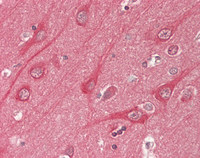
| Tissue Location | Brain-specific. |
Research Areas
Citations (0)

Thousands of laboratories across the world have published research that depended on the performance of antibodies from Abcepta to advance their research. Check out links to articles that cite our products in major peer-reviewed journals, organized by research category.
Submit your citation using an Abcepta antibody to
info@abcepta.com, and receive a free "I Love Antibodies" mug.
info@abcepta.com, and receive a free "I Love Antibodies" mug.

Application Protocols
Provided below are standard protocols that you may find useful for product applications.
Abcepta welcomes feedback from its customers.
If you have used an Abcepta product and would like to share how it has performed, please click on the "Submit Review" button and provide the requested information. Our staff will examine and post your review and contact you if needed.
If you have any additional inquiries please email technical services at tech@abcepta.com.
$ 467.50
Cat# ALS17330
Ordering Information
United States
AlbaniaAustraliaAustriaBelgiumBosnia & HerzegovinaBrazilBulgariaCanadaCentral AmericaChinaCroatiaCyprusCzech RepublicDenmarkEstoniaFinlandFranceGermanyGreeceHong KongHungaryIcelandIndiaIndonesiaIrelandIsraelItalyJapanLatviaLithuaniaLuxembourgMacedoniaMalaysiaMaltaNetherlandsNew ZealandNorwayPakistanPolandPortugalRomaniaSerbiaSingaporeSlovakiaSloveniaSouth AfricaSouth KoreaSpainSwedenSwitzerlandTaiwanTurkeyUnited KingdomUnited StatesVietnamWorldwideOthers
USA Headquarters
(888) 735-7227 / (858) 622-0099 or (858) 875-1900
Other Products
Shipping Information
Domestic orders (in stock items)
Shipped out the same day. Orders placed after 1 PM (PST) will ship out the next business day.
International orders
Contact your local distributors
