


 Foundational characteristics of cancer include proliferation, angiogenesis, migration, evasion of apoptosis, and cellular immortality. Find key markers for these cellular processes and antibodies to detect them.
 The SUMOplot™ Analysis Program predicts and scores sumoylation sites in your protein. SUMOylation is a post-translational modification involved in various cellular processes, such as nuclear-cytosolic transport, transcriptional regulation, apoptosis, protein stability, response to stress, and progression through the cell cycle.
 The Autophagy Receptor Motif Plotter predicts and scores autophagy receptor binding sites in your protein. Identifying proteins connected to this pathway is critical to understanding the role of autophagy in physiological as well as pathological processes such as development, differentiation, neurodegenerative diseases, stress, infection, and cancer.
GFP Tag Antibody
Purified Mouse Monoclonal Antibody (Mab)
- SPECIFICATION
- CITATIONS: 26
- PROTOCOLS
- BACKGROUND

Application
| WB, IF, E |
|---|---|
| Primary Accession | C5MKY7 |
| Reactivity | Human |
| Host | Mouse |
| Clonality | Monoclonal |
| Isotype | Mouse IgG1 |
| Clone/Animal Names | 168AT1211 |
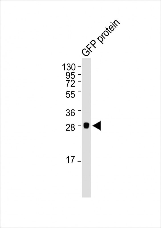
| Calculated MW | 27 k KDa |
| Target/Specificity | Purified His-tagged GFP protein was used to produced this monoclonal antibody. |
|---|---|
| Dilution | IF~~1:25 WB~~1:20000 |
| Format | Purified monoclonal antibody supplied in PBS with 0.09% (W/V) sodium azide. This antibody is purified through a protein G column, followed by dialysis against PBS. |
| Storage | Maintain refrigerated at 2-8°C for up to 2 weeks. For long term storage store at -20°C in small aliquots to prevent freeze-thaw cycles. |
| Precautions | GFP Tag Antibody is for research use only and not for use in diagnostic or therapeutic procedures. |

Provided below are standard protocols that you may find useful for product applications.
Background
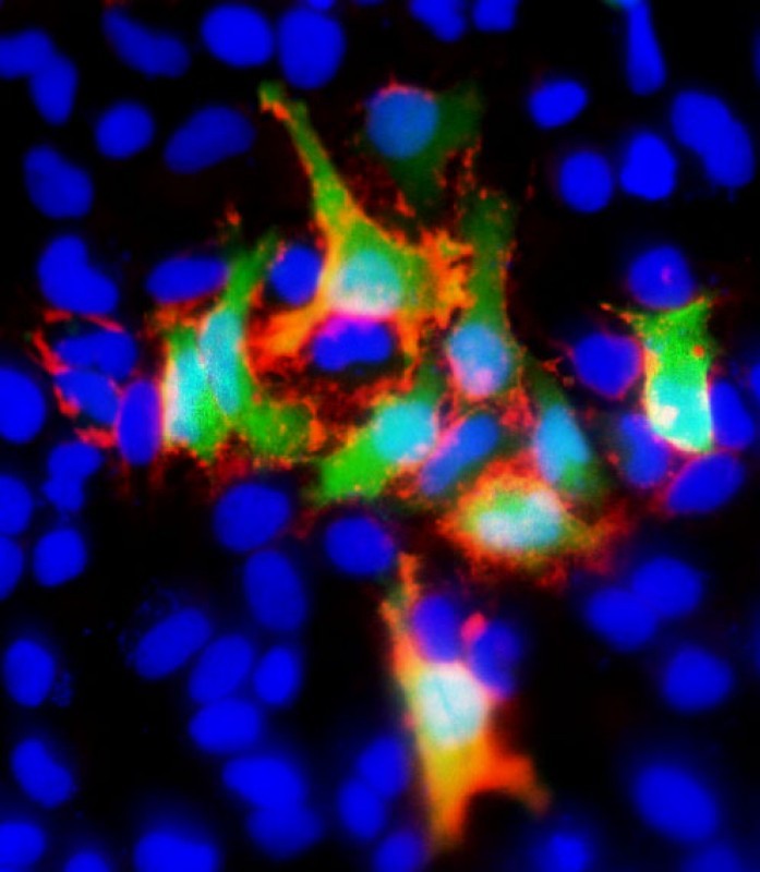
Green fluorescent protein (GFP), originally isolated from the jellyfish Aequorea victoria, is one of the best visual reporters for monitoring gene expression in vivo and in situ. GFP is a also convenient marker for use in flow cytometry because it eliminates the need to incubate with a secondary reagent (such as dyes or antibodies) for detection. However, anti-GFP antibody is also widely used for co-immunipreciapitation, co-localization or western blotting for the confirmation of specificity when a GFP fusion protein is expressed in cells. Abgent's anti-GFP monoclonal antibody provides a simple solution to detect the expression of a GFP-tagged protein in cells. Because of its ability to spontaneously generate its own fluorophore, the green fluorescent protein (GFP) from the jellyfish Aequorea victoria is used extensively as a fluorescent marker in molecular and cell biology. The yellow fluorescent proteins (YFPs) have the longest wavelength emissions of all GFP variants examined to date. This shift in the spectrum is the result of a T203Y substitution (single-letter amino acid code), a mutation rationally designed on the basis of the X-ray structure of GFP S65T. Abgent's anti-GFP monoclonal antibody can detect both GFP and YFP but not BFP (Blue fluorescent protein) by western blotting.
References
Ward, W. W., et al.(1980) Photochem. Photobiol. 31:611
If you have used an Abcepta product and would like to share how it has performed, please click on the "Submit Review" button and provide the requested information. Our staff will examine and post your review and contact you if needed.
If you have any additional inquiries please email technical services at tech@abcepta.com.
Ordering Information
Other Products
Shipping Information
