


 Foundational characteristics of cancer include proliferation, angiogenesis, migration, evasion of apoptosis, and cellular immortality. Find key markers for these cellular processes and antibodies to detect them.
 The SUMOplot™ Analysis Program predicts and scores sumoylation sites in your protein. SUMOylation is a post-translational modification involved in various cellular processes, such as nuclear-cytosolic transport, transcriptional regulation, apoptosis, protein stability, response to stress, and progression through the cell cycle.
 The Autophagy Receptor Motif Plotter predicts and scores autophagy receptor binding sites in your protein. Identifying proteins connected to this pathway is critical to understanding the role of autophagy in physiological as well as pathological processes such as development, differentiation, neurodegenerative diseases, stress, infection, and cancer.
NTRK2 Antibody
Purified Mouse Monoclonal Antibody (Mab)

- SPECIFICATION
- CITATIONS
- PROTOCOLS
- BACKGROUND

Application
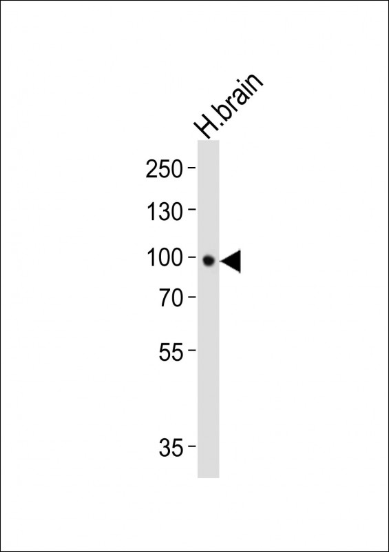
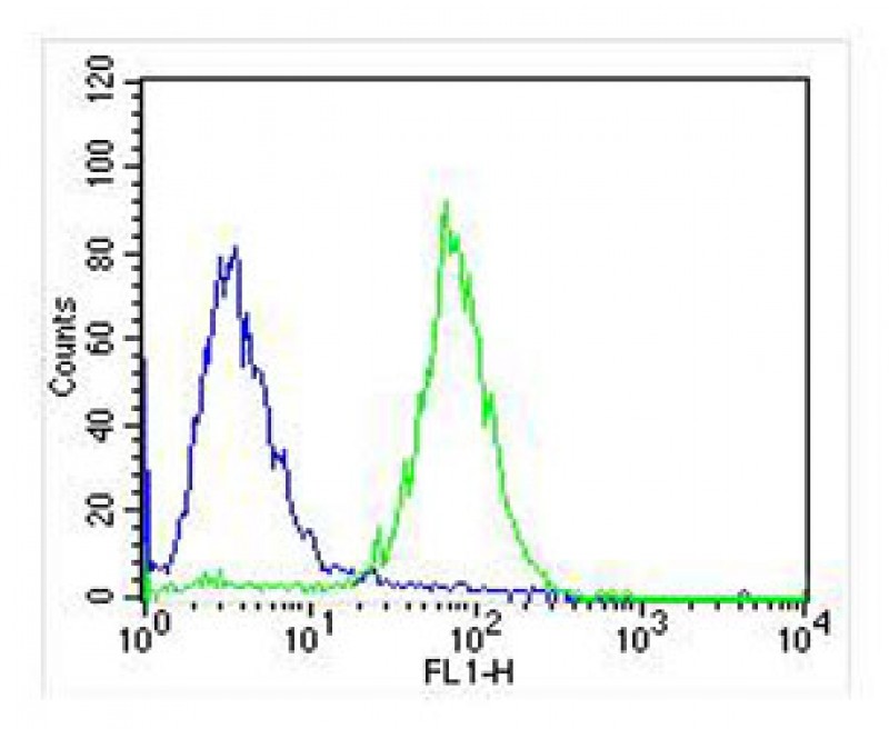
| WB, FC, IHC-P, E |
|---|---|
| Primary Accession | Q16620 |
| Reactivity | Human |
| Host | Mouse |
| Clonality | monoclonal |
| Isotype | IgG1,k |
| Clone/Animal Names | 1446CT494.85.83.49 |
| Calculated MW | 91999 Da |
| Gene ID | 4915 |
|---|---|
| Other Names | BDNF/NT-3 growth factors receptor, GP145-TrkB, Trk-B, Neurotrophic tyrosine kinase receptor type 2, TrkB tyrosine kinase, Tropomyosin-related kinase B, NTRK2, TRKB |
| Target/Specificity | This NTRK2 antibody is generated from a mouse immunized with a recombinant protein. |
| Dilution | WB~~1:1000 IHC-P~~1:25 FC~~1:25 |
| Format | Purified monoclonal antibody supplied in PBS with 0.09% (W/V) sodium azide. This antibody is purified through a protein G column, followed by dialysis against PBS. |
| Storage | Maintain refrigerated at 2-8°C for up to 2 weeks. For long term storage store at -20°C in small aliquots to prevent freeze-thaw cycles. |
| Precautions | NTRK2 Antibody is for research use only and not for use in diagnostic or therapeutic procedures. |
| Name | NTRK2 |
|---|---|
| Synonyms | TRKB |
| Function | Receptor tyrosine kinase involved in the development and the maturation of the central and the peripheral nervous systems through regulation of neuron survival, proliferation, migration, differentiation, and synapse formation and plasticity (By similarity). Receptor for BDNF/brain-derived neurotrophic factor and NTF4/neurotrophin-4. Alternatively can also bind NTF3/neurotrophin-3 which is less efficient in activating the receptor but regulates neuron survival through NTRK2 (PubMed:7574684, PubMed:15494731). Upon ligand- binding, undergoes homodimerization, autophosphorylation and activation (PubMed:15494731). Recruits, phosphorylates and/or activates several downstream effectors including SHC1, FRS2, SH2B1, SH2B2 and PLCG1 that regulate distinct overlapping signaling cascades. Through SHC1, FRS2, SH2B1, SH2B2 activates the GRB2-Ras-MAPK cascade that regulates for instance neuronal differentiation including neurite outgrowth. Through the same effectors controls the Ras-PI3 kinase-AKT1 signaling cascade that mainly regulates growth and survival. Through PLCG1 and the downstream protein kinase C-regulated pathways controls synaptic plasticity. Thereby, plays a role in learning and memory by regulating both short term synaptic function and long-term potentiation. PLCG1 also leads to NF-Kappa-B activation and the transcription of genes involved in cell survival. Hence, it is able to suppress anoikis, the apoptosis resulting from loss of cell-matrix interactions. May also play a role in neutrophin-dependent calcium signaling in glial cells and mediate communication between neurons and glia. |
| Cellular Location | Cell membrane; Single-pass type I membrane protein. Endosome membrane {ECO:0000250|UniProtKB:P15209}; Single-pass type I membrane protein {ECO:0000250|UniProtKB:P15209}. Early endosome membrane {ECO:0000250|UniProtKB:P15209}. Cell projection, axon {ECO:0000250|UniProtKB:Q63604}. Cell projection, dendrite {ECO:0000250|UniProtKB:Q63604}. Cytoplasm, perinuclear region {ECO:0000250|UniProtKB:Q63604}. Postsynaptic density {ECO:0000250|UniProtKB:P15209}. Note=Internalized to endosomes upon ligand-binding. {ECO:0000250|UniProtKB:P15209} |
| Tissue Location | Isoform TrkB is expressed in the central and peripheral nervous system. In the central nervous system (CNS), expression is observed in the cerebral cortex, hippocampus, thalamus, choroid plexus, granular layer of the cerebellum, brain stem, and spinal cord. In the peripheral nervous system, it is expressed in many cranial ganglia, the ophthalmic nerve, the vestibular system, multiple facial structures, the submaxillary glands, and dorsal root ganglia Isoform TrkB-T1 is mainly expressed in the brain but also detected in other tissues including pancreas, kidney and heart. Isoform TrkB-T-Shc is predominantly expressed in the brain. |

Thousands of laboratories across the world have published research that depended on the performance of antibodies from Abcepta to advance their research. Check out links to articles that cite our products in major peer-reviewed journals, organized by research category.
info@abcepta.com, and receive a free "I Love Antibodies" mug.

Provided below are standard protocols that you may find useful for product applications.
Background
Receptor tyrosine kinase involved in the development and the maturation of the central and the peripheral nervous systems through regulation of neuron survival, proliferation, migration, differentiation, and synapse formation and plasticity. Receptor for BDNF/brain-derived neurotrophic factor and NTF4/neurotrophin- 4. Alternatively can also bind NTF3/neurotrophin-3 which is less efficient in activating the receptor but regulates neuron survival through NTRK2. Upon ligand-binding, undergoes homodimerization, autophosphorylation and activation. Recruits, phosphorylates and/or activates several downstream effectors including SHC1, FRS2, SH2B1, SH2B2 and PLCG1 that regulate distinct overlapping signaling cascades. Through SHC1, FRS2, SH2B1, SH2B2 activates the GRB2-Ras-MAPK cascade that regulates for instance neuronal differentiation including neurite outgrowth. Through the same effectors controls the Ras-PI3 kinase-AKT1 signaling cascade that mainly regulates growth and survival. Through PLCG1 and the downstream protein kinase C-regulated pathways controls synaptic plasticity. Thereby, plays a role in learning and memory by regulating both short term synaptic function and long-term potentiation. PLCG1 also leads to NF-Kappa-B activation and the transcription of genes involved in cell survival. Hence, it is able to suppress anoikis, the apoptosis resulting from loss of cell-matrix interactions. May also play a role in neutrophin- dependent calcium signaling in glial cells and mediate communication between neurons and glia.
References
Nakagawara A.,et al.Genomics 25:538-546(1995).
Shelton D.L.,et al.J. Neurosci. 15:477-491(1995).
Allen S.J.,et al.Neuroscience 60:825-834(1994).
Stoilov P.,et al.Biochem. Biophys. Res. Commun. 290:1054-1065(2002).
Steinbeck J.A.,et al.Submitted (MAY-2002) to the EMBL/GenBank/DDBJ databases.
If you have used an Abcepta product and would like to share how it has performed, please click on the "Submit Review" button and provide the requested information. Our staff will examine and post your review and contact you if needed.
If you have any additional inquiries please email technical services at tech@abcepta.com.
Ordering Information
Other Products
Shipping Information
