


 Foundational characteristics of cancer include proliferation, angiogenesis, migration, evasion of apoptosis, and cellular immortality. Find key markers for these cellular processes and antibodies to detect them.
 The SUMOplot™ Analysis Program predicts and scores sumoylation sites in your protein. SUMOylation is a post-translational modification involved in various cellular processes, such as nuclear-cytosolic transport, transcriptional regulation, apoptosis, protein stability, response to stress, and progression through the cell cycle.
 The Autophagy Receptor Motif Plotter predicts and scores autophagy receptor binding sites in your protein. Identifying proteins connected to this pathway is critical to understanding the role of autophagy in physiological as well as pathological processes such as development, differentiation, neurodegenerative diseases, stress, infection, and cancer.
IRF3 Antibody
Purified Mouse Monoclonal Antibody (Mab)
- SPECIFICATION
- CITATIONS
- PROTOCOLS
- BACKGROUND

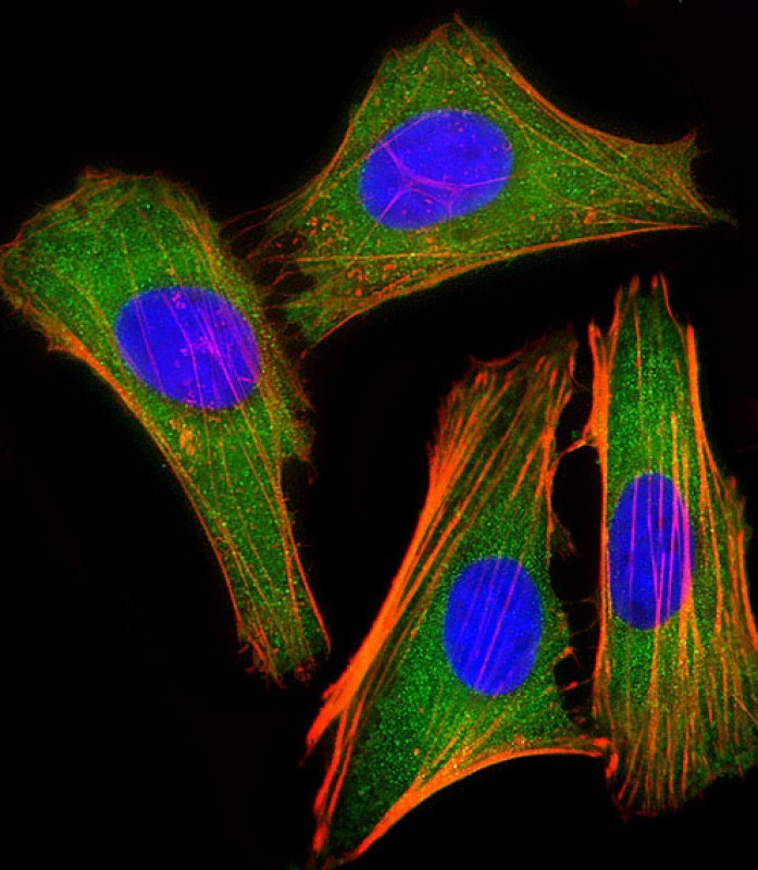
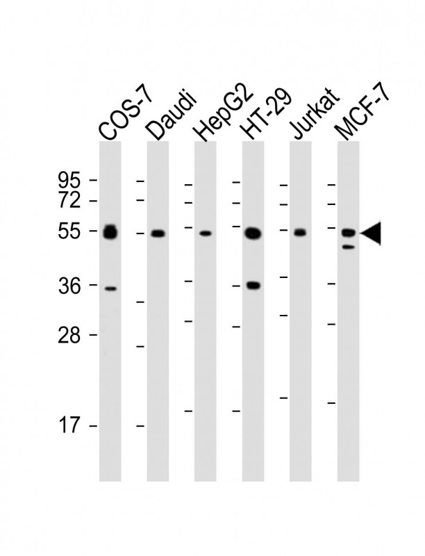
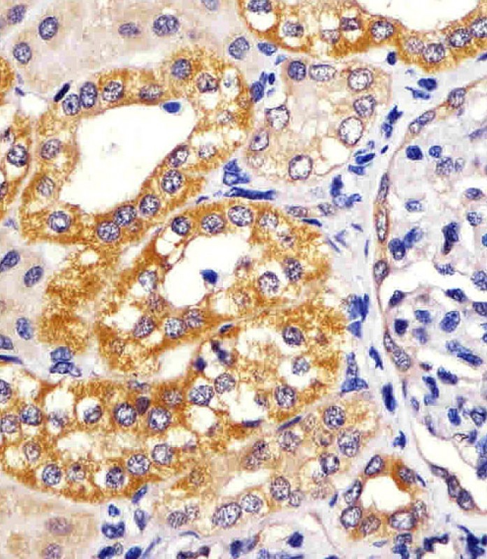
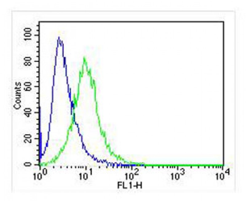
Application
| WB, IHC-P, FC, IF, E |
|---|---|
| Primary Accession | Q14653 |
| Reactivity | Human, African Green Monkey |
| Host | Mouse |
| Clonality | monoclonal |
| Isotype | IgG1,k |
| Clone/Animal Names | 1522CT766.58.24 |
| Calculated MW | 47219 Da |
| Gene ID | 3661 |
|---|---|
| Other Names | Interferon regulatory factor 3, IRF-3, IRF3 |
| Target/Specificity | This IRF3 antibody is generated from a mouse immunized with a recombinant protein. |
| Dilution | IF~~1:25 WB~~1:2000 IHC-P~~1:25 FC~~1:25 |
| Format | Purified monoclonal antibody supplied in PBS with 0.09% (W/V) sodium azide. This antibody is purified through a protein G column, followed by dialysis against PBS. |
| Storage | Maintain refrigerated at 2-8°C for up to 2 weeks. For long term storage store at -20°C in small aliquots to prevent freeze-thaw cycles. |
| Precautions | IRF3 Antibody is for research use only and not for use in diagnostic or therapeutic procedures. |
| Name | IRF3 {ECO:0000303|PubMed:9803267, ECO:0000312|HGNC:HGNC:6118} |
|---|---|
| Function | Key transcriptional regulator of type I interferon (IFN)- dependent immune responses which plays a critical role in the innate immune response against DNA and RNA viruses (PubMed:8524823, PubMed:22394562, PubMed:25636800, PubMed:27302953, PubMed:24049179, PubMed:31340999, PubMed:36603579). Regulates the transcription of type I IFN genes (IFN-alpha and IFN-beta) and IFN-stimulated genes (ISG) by binding to an interferon-stimulated response element (ISRE) in their promoters (PubMed:8524823, PubMed:11846977, PubMed:16846591, PubMed:16979567, PubMed:20049431, PubMed:36603579, PubMed:32972995). Acts as a more potent activator of the IFN-beta (IFNB) gene than the IFN-alpha (IFNA) gene and plays a critical role in both the early and late phases of the IFNA/B gene induction (PubMed:16846591, PubMed:16979567, PubMed:20049431, PubMed:36603579). Found in an inactive form in the cytoplasm of uninfected cells and following viral infection, double-stranded RNA (dsRNA), or toll-like receptor (TLR) signaling, is phosphorylated by IKBKE and TBK1 kinases (PubMed:22394562, PubMed:25636800, PubMed:36603579, PubMed:27302953). This induces a conformational change, leading to its dimerization and nuclear localization and association with CREB binding protein (CREBBP) to form dsRNA-activated factor 1 (DRAF1), a complex which activates the transcription of the type I IFN and ISG genes (PubMed:16154084, PubMed:27302953, PubMed:33440148, PubMed:36603579). Can activate distinct gene expression programs in macrophages and can induce significant apoptosis in primary macrophages (PubMed:16846591). In response to Sendai virus infection, is recruited by TOMM70:HSP90AA1 to mitochondrion and forms an apoptosis complex TOMM70:HSP90AA1:IRF3:BAX inducing apoptosis (PubMed:25609812). Key transcription factor regulating the IFN response during SARS-CoV-2 infection (PubMed:33440148). |
| Cellular Location | Cytoplasm. Nucleus Mitochondrion. Note=Shuttles between cytoplasmic and nuclear compartments, with export being the prevailing effect (PubMed:10805757, PubMed:35922005). When activated, IRF3 interaction with CREBBP prevents its export to the cytoplasm (PubMed:10805757). Recruited to mitochondria via TOMM70:HSP90AA1 upon Sendai virus infection (PubMed:25609812). |
| Tissue Location | Expressed constitutively in a variety of tissues. |

Thousands of laboratories across the world have published research that depended on the performance of antibodies from Abcepta to advance their research. Check out links to articles that cite our products in major peer-reviewed journals, organized by research category.
info@abcepta.com, and receive a free "I Love Antibodies" mug.

Provided below are standard protocols that you may find useful for product applications.
Background
Key transcriptional regulator of type I interferon (IFN)-dependent immune responses which plays a critical role in the innate immune response against DNA and RNA viruses. Regulates the transcription of type I IFN genes (IFN-alpha and IFN-beta) and IFN-stimulated genes (ISG) by binding to an interferon-stimulated response element (ISRE) in their promoters. Acts as a more potent activator of the IFN-beta (IFNB) gene than the IFN-alpha (IFNA) gene and plays a critical role in both the early and late phases of the IFNA/B gene induction. Found in an inactive form in the cytoplasm of uninfected cells and following viral infection, double-stranded RNA (dsRNA), or toll-like receptor (TLR) signaling, is phosphorylated by IKBKE and TBK1 kinases. This induces a conformational change, leading to its dimerization and nuclear localization and association with CREB binding protein (CREBBP) to form dsRNA-activated factor 1 (DRAF1), a complex which activates the transcription of the type I IFN and ISG genes. Can activate distinct gene expression programs in macrophages and can induce significant apoptosis in primary macrophages.
References
Au W.W.-C.,et al.Proc. Natl. Acad. Sci. U.S.A. 92:11657-11661(1995).
Tabata Y.,et al.Submitted (FEB-2003) to the EMBL/GenBank/DDBJ databases.
Ota T.,et al.Nat. Genet. 36:40-45(2004).
Grimwood J.,et al.Nature 428:529-535(2004).
Mural R.J.,et al.Submitted (JUL-2005) to the EMBL/GenBank/DDBJ databases.
If you have used an Abcepta product and would like to share how it has performed, please click on the "Submit Review" button and provide the requested information. Our staff will examine and post your review and contact you if needed.
If you have any additional inquiries please email technical services at tech@abcepta.com.
Ordering Information
Other Products
Shipping Information
