


 Foundational characteristics of cancer include proliferation, angiogenesis, migration, evasion of apoptosis, and cellular immortality. Find key markers for these cellular processes and antibodies to detect them.
 The SUMOplot™ Analysis Program predicts and scores sumoylation sites in your protein. SUMOylation is a post-translational modification involved in various cellular processes, such as nuclear-cytosolic transport, transcriptional regulation, apoptosis, protein stability, response to stress, and progression through the cell cycle.
 The Autophagy Receptor Motif Plotter predicts and scores autophagy receptor binding sites in your protein. Identifying proteins connected to this pathway is critical to understanding the role of autophagy in physiological as well as pathological processes such as development, differentiation, neurodegenerative diseases, stress, infection, and cancer.
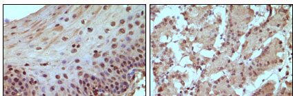
RB Antibody
Purified Mouse Monoclonal Antibody
- SPECIFICATION
- CITATIONS
- PROTOCOLS
- BACKGROUND

Application
| IHC, E |
|---|---|
| Primary Accession | P06400 |
| Reactivity | Human |
| Host | Mouse |
| Clonality | Monoclonal |
| Clone Names | 7E4B8 |
| Isotype | IgG1 |
| Calculated MW | 106159 Da |
| Description | The Rb protein regulates differentiation, apoptosis, and cell cycle control by coordinating the cell cycle at G1-S with transcriptional machinery. During G1, cyclin D-dependent kinase-mediated phosphorylation of Rb at Ser-795 marks the conversion of Rb from a transcriptionally repressive, hypophosphorylated state to an inactive, phosphorylated state, which may be sustained through mitosis by differential phosphorylation of up to 16 putative serine or threonine residues. Pediatric cancer retinoblastoma and the formation of other human tumors can be attributed to mutations in the retinoblastoma tumor suppressor gene(Rb). |
| Immunogen | Purified recombinant fragment of human RB expressed in E. Coli. |
| Formulation | Ascitic fluid containing 0.03% sodium azide. |
| Gene ID | 5925 |
|---|---|
| Other Names | Retinoblastoma-associated protein, p105-Rb, pRb, Rb, pp110, RB1 |
| Dilution | IHC~~1/200 - 1/1000 |
| Storage | Maintain refrigerated at 2-8°C for up to 6 months. For long term storage store at -20°C in small aliquots to prevent freeze-thaw cycles. |
| Precautions | RB Antibody is for research use only and not for use in diagnostic or therapeutic procedures. |
| Name | RB1 |
|---|---|
| Function | Tumor suppressor that is a key regulator of the G1/S transition of the cell cycle (PubMed:10499802). The hypophosphorylated form binds transcription regulators of the E2F family, preventing transcription of E2F-responsive genes (PubMed:10499802). Both physically blocks E2Fs transactivating domain and recruits chromatin- modifying enzymes that actively repress transcription (PubMed:10499802). Cyclin and CDK-dependent phosphorylation of RB1 induces its dissociation from E2Fs, thereby activating transcription of E2F responsive genes and triggering entry into S phase (PubMed:10499802). RB1 also promotes the G0-G1 transition upon phosphorylation and activation by CDK3/cyclin-C (PubMed:15084261). Directly involved in heterochromatin formation by maintaining overall chromatin structure and, in particular, that of constitutive heterochromatin by stabilizing histone methylation. Recruits and targets histone methyltransferases SUV39H1, KMT5B and KMT5C, leading to epigenetic transcriptional repression. Controls histone H4 'Lys-20' trimethylation. Inhibits the intrinsic kinase activity of TAF1. Mediates transcriptional repression by SMARCA4/BRG1 by recruiting a histone deacetylase (HDAC) complex to the c-FOS promoter. In resting neurons, transcription of the c-FOS promoter is inhibited by BRG1- dependent recruitment of a phospho-RB1-HDAC1 repressor complex. Upon calcium influx, RB1 is dephosphorylated by calcineurin, which leads to release of the repressor complex (By similarity). |
| Cellular Location | Nucleus. Note=During keratinocyte differentiation, acetylation by KAT2B/PCAF is required for nuclear localization. |
| Tissue Location | Expressed in the retina. Expressed in foreskin keratinocytes (at protein level) (PubMed:20940255) |
Research Areas
Citations (0)

Thousands of laboratories across the world have published research that depended on the performance of antibodies from Abcepta to advance their research. Check out links to articles that cite our products in major peer-reviewed journals, organized by research category.
Submit your citation using an Abcepta antibody to
info@abcepta.com, and receive a free "I Love Antibodies" mug.
info@abcepta.com, and receive a free "I Love Antibodies" mug.

Application Protocols
Provided below are standard protocols that you may find useful for product applications.
References
1. Oncogene 19: 562-570. 2. Cell 81: 323-330.
Abcepta welcomes feedback from its customers.
If you have used an Abcepta product and would like to share how it has performed, please click on the "Submit Review" button and provide the requested information. Our staff will examine and post your review and contact you if needed.
If you have any additional inquiries please email technical services at tech@abcepta.com.
$ 300.00
Cat# AO1031a
Ordering Information
United States
AlbaniaAustraliaAustriaBelgiumBosnia & HerzegovinaBrazilBulgariaCanadaCentral AmericaChinaCroatiaCyprusCzech RepublicDenmarkEstoniaFinlandFranceGermanyGreeceHong KongHungaryIcelandIndiaIndonesiaIrelandIsraelItalyJapanLatviaLithuaniaLuxembourgMacedoniaMalaysiaMaltaNetherlandsNew ZealandNorwayPakistanPolandPortugalRomaniaSerbiaSingaporeSlovakiaSloveniaSouth AfricaSouth KoreaSpainSwedenSwitzerlandTaiwanTurkeyUnited KingdomUnited StatesVietnamWorldwideOthers
USA Headquarters
(888) 735-7227 / (858) 622-0099 or (858) 875-1900
Other Products
Shipping Information
Domestic orders (in stock items)
Shipped out the same day. Orders placed after 1 PM (PST) will ship out the next business day.
International orders
Contact your local distributors
