


 Foundational characteristics of cancer include proliferation, angiogenesis, migration, evasion of apoptosis, and cellular immortality. Find key markers for these cellular processes and antibodies to detect them.
 The SUMOplot™ Analysis Program predicts and scores sumoylation sites in your protein. SUMOylation is a post-translational modification involved in various cellular processes, such as nuclear-cytosolic transport, transcriptional regulation, apoptosis, protein stability, response to stress, and progression through the cell cycle.
 The Autophagy Receptor Motif Plotter predicts and scores autophagy receptor binding sites in your protein. Identifying proteins connected to this pathway is critical to understanding the role of autophagy in physiological as well as pathological processes such as development, differentiation, neurodegenerative diseases, stress, infection, and cancer.
EphB4 Antibody
Purified Mouse Monoclonal Antibody
- SPECIFICATION
- CITATIONS
- PROTOCOLS
- BACKGROUND

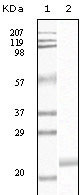
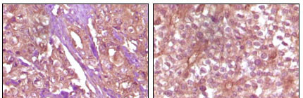
Application
| WB, IHC, E |
|---|---|
| Primary Accession | P54760 |
| Reactivity | Human |
| Host | Mouse |
| Clonality | Monoclonal |
| Clone Names | 5B8F7 |
| Isotype | IgG2a |
| Calculated MW | 108270 Da |
| Description | EPH receptor B4 (EphB4), with 987-amino acid protein (about 108kDa), belongs to the ephrin receptor subfamily of the protein-tyrosine kinase family. The Eph receptor tyrosine kinases and their ligands, the ephrins, regulate numerous biological processes in developing and adult tissues and have been implicated in cancer progression and in pathological forms of angiogenesis. EphB4 acts as a negative regulator of blood vessel branching and vascular network formation, switching the vascularization program from sprouting angiogenesis to circumferential vessel growth. EphB4 and its ligand ephrinB2 express in several kinds of tumor cells and correlate with tumorigenesis. EphB4 is thus a potential candidate as a predictor of disease outcome in several kinds of tumor and as target for novel therapy. |
| Immunogen | Purified recombinant fragment of EphB4 expressed in E. Coli. |
| Formulation | Ascitic fluid containing 0.03% sodium azide. |
| Gene ID | 2050 |
|---|---|
| Other Names | Ephrin type-B receptor 4, 2.7.10.1, Hepatoma transmembrane kinase, Tyrosine-protein kinase TYRO11, EPHB4, HTK, MYK1, TYRO11 |
| Dilution | WB~~1/500 - 1/2000 IHC~~1/200 - 1/1000 |
| Storage | Maintain refrigerated at 2-8°C for up to 6 months. For long term storage store at -20°C in small aliquots to prevent freeze-thaw cycles. |
| Precautions | EphB4 Antibody is for research use only and not for use in diagnostic or therapeutic procedures. |
| Name | EPHB4 |
|---|---|
| Synonyms | HTK, MYK1, TYRO11 |
| Function | Receptor tyrosine kinase which binds promiscuously transmembrane ephrin-B family ligands residing on adjacent cells, leading to contact-dependent bidirectional signaling into neighboring cells. The signaling pathway downstream of the receptor is referred to as forward signaling while the signaling pathway downstream of the ephrin ligand is referred to as reverse signaling. Together with its cognate ligand/functional ligand EFNB2 it is involved in the regulation of cell adhesion and migration, and plays a central role in heart morphogenesis, angiogenesis and blood vessel remodeling and permeability. EPHB4-mediated forward signaling controls cellular repulsion and segregation from EFNB2-expressing cells. |
| Cellular Location | Cell membrane; Single-pass type I membrane protein |
| Tissue Location | Abundantly expressed in placenta but also detected in kidney, liver, lung, pancreas, skeletal muscle and heart. Expressed in primitive and myeloid, but not lymphoid, hematopoietic cells. Also observed in cell lines derived from liver, breast, colon, lung, melanocyte and cervix. |

Thousands of laboratories across the world have published research that depended on the performance of antibodies from Abcepta to advance their research. Check out links to articles that cite our products in major peer-reviewed journals, organized by research category.
info@abcepta.com, and receive a free "I Love Antibodies" mug.

Provided below are standard protocols that you may find useful for product applications.
References
1. J. Chrencik, A. Brooun, M. Recht. Structure. 2006 Feb;14(2):321-30. 2. Qinghua WU, Zhenhe SUO,Bjon RISBERG. Pathol Oncol Res. 2004;10(1):26-33.
If you have used an Abcepta product and would like to share how it has performed, please click on the "Submit Review" button and provide the requested information. Our staff will examine and post your review and contact you if needed.
If you have any additional inquiries please email technical services at tech@abcepta.com.
Ordering Information
Other Products
Shipping Information
