


 Foundational characteristics of cancer include proliferation, angiogenesis, migration, evasion of apoptosis, and cellular immortality. Find key markers for these cellular processes and antibodies to detect them.
 The SUMOplot™ Analysis Program predicts and scores sumoylation sites in your protein. SUMOylation is a post-translational modification involved in various cellular processes, such as nuclear-cytosolic transport, transcriptional regulation, apoptosis, protein stability, response to stress, and progression through the cell cycle.
 The Autophagy Receptor Motif Plotter predicts and scores autophagy receptor binding sites in your protein. Identifying proteins connected to this pathway is critical to understanding the role of autophagy in physiological as well as pathological processes such as development, differentiation, neurodegenerative diseases, stress, infection, and cancer.
STYK1 Antibody
Purified Mouse Monoclonal Antibody
- SPECIFICATION
- CITATIONS
- PROTOCOLS
- BACKGROUND

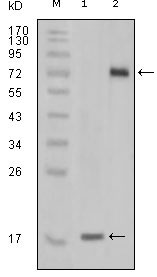
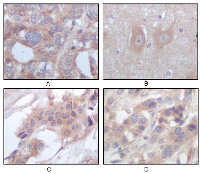
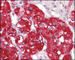
Application
| WB, IHC, E |
|---|---|
| Primary Accession | Q6J9G0 |
| Reactivity | Human |
| Host | Mouse |
| Clonality | Monoclonal |
| Clone Names | 2H2F10 |
| Isotype | IgG1 |
| Calculated MW | 47577 Da |
| Description | Protein kinases (PKs) represent a well studied but most diverse protein superfamily. The covalent, reversible linkage of phosphate to serine, threonine, and tyrosine residues of substrate proteins by protein kinases is probably ubiquitous cellular mechanism for regulation of physiological processes. It is known to us that most signaling pathways impinge at some point on protein kinases. Here we report a human putative receptor protein kinase cDNA STYK1. The STYK1 cDNA is 2749 base pairs in length and contains an open reading frame encoding 422 amino acids. The STYK1 gene is mapped to human chromosome 12p13 and 11 exons were found. RT-PCR showed that STYK1 is widely expressed in human tissues. |
| Immunogen | Purified recombinant fragment of STYK1 expressed in E. Coli. |
| Formulation | Ascitic fluid containing 0.03% sodium azide. |
| Gene ID | 55359 |
|---|---|
| Other Names | Tyrosine-protein kinase STYK1, 2.7.10.2, Novel oncogene with kinase domain, Protein PK-unique, Serine/threonine/tyrosine kinase 1, STYK1, NOK |
| Dilution | WB~~1/500 - 1/2000 IHC~~1/500 - 1/2000 |
| Storage | Maintain refrigerated at 2-8°C for up to 6 months. For long term storage store at -20°C in small aliquots to prevent freeze-thaw cycles. |
| Precautions | STYK1 Antibody is for research use only and not for use in diagnostic or therapeutic procedures. |
| Name | STYK1 |
|---|---|
| Synonyms | NOK |
| Function | Probable tyrosine protein-kinase, which has strong transforming capabilities on a variety of cell lines. When overexpressed, it can also induce tumor cell invasion as well as metastasis in distant organs. May act by activating both MAP kinase and phosphatidylinositol 3'-kinases (PI3K) pathways (By similarity). |
| Cellular Location | Membrane; Single-pass membrane protein |
| Tissue Location | Widely expressed. Highly expressed in brain, placenta and prostate. Expressed in tumor cells such as hepatoma cells L-02, cervix carcinoma cells HeLa, ovary cancer cells Ho8910 and chronic myelogenous leukemia cells K-562, but not in other tumor cells such as epidermoid carcinoma (A-431). Undetectable in most normal lung tissues, widely expressed in lung cancers |

Thousands of laboratories across the world have published research that depended on the performance of antibodies from Abcepta to advance their research. Check out links to articles that cite our products in major peer-reviewed journals, organized by research category.
info@abcepta.com, and receive a free "I Love Antibodies" mug.

Provided below are standard protocols that you may find useful for product applications.
References
1. Liu L, Yu XZ and Li TS, et al. Mol Biol Rep. 2003, Jun, 30(2):91-6. 2. Moriai R. , Kobayashi D.and Amachika T. , et al. Mol Biol Rep. 2007, Apr, 6.
If you have used an Abcepta product and would like to share how it has performed, please click on the "Submit Review" button and provide the requested information. Our staff will examine and post your review and contact you if needed.
If you have any additional inquiries please email technical services at tech@abcepta.com.
Ordering Information
Other Products
Shipping Information
