


 Foundational characteristics of cancer include proliferation, angiogenesis, migration, evasion of apoptosis, and cellular immortality. Find key markers for these cellular processes and antibodies to detect them.
 The SUMOplot™ Analysis Program predicts and scores sumoylation sites in your protein. SUMOylation is a post-translational modification involved in various cellular processes, such as nuclear-cytosolic transport, transcriptional regulation, apoptosis, protein stability, response to stress, and progression through the cell cycle.
 The Autophagy Receptor Motif Plotter predicts and scores autophagy receptor binding sites in your protein. Identifying proteins connected to this pathway is critical to understanding the role of autophagy in physiological as well as pathological processes such as development, differentiation, neurodegenerative diseases, stress, infection, and cancer.
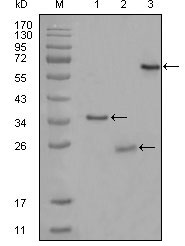
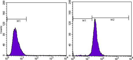
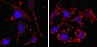
CD33 Antibody
Purified Mouse Monoclonal Antibody

- SPECIFICATION
- CITATIONS: 5
- PROTOCOLS
- BACKGROUND

Application
| WB, E |
|---|---|
| Primary Accession | P20138 |
| Reactivity | Human |
| Host | Mouse |
| Clonality | Monoclonal |
| Clone Names | 2B7C12 |
| Isotype | IgG1 |
| Calculated MW | 39825 Da |
| Description | CD33 is found on granulocyte and macrophage precursors in the bone marrow, but is not on pluripotent stem cells. The protein is also expressed on, and is a useful marker for, peripheral monocytes. It is also useful for distinguishing myelogenous leukaemia cells from lymphoid or erythroid leukaemias. |
| Immunogen | Purified recombinant fragment of CD33 (48-258) expressed in E. Coli. |
| Formulation | Ascitic fluid containing 0.03% sodium azide. |
| Gene ID | 945 |
|---|---|
| Other Names | Myeloid cell surface antigen CD33, Sialic acid-binding Ig-like lectin 3, Siglec-3, gp67, CD33, CD33, SIGLEC3 |
| Dilution | WB~~1/500 - 1/2000 FC~~1:200~~400 ICC~~1:200~~1000 IF~~1:200~1000. |
| Storage | Maintain refrigerated at 2-8°C for up to 6 months. For long term storage store at -20°C in small aliquots to prevent freeze-thaw cycles. |
| Precautions | CD33 Antibody is for research use only and not for use in diagnostic or therapeutic procedures. |
| Name | CD33 |
|---|---|
| Synonyms | SIGLEC3 |
| Function | Sialic-acid-binding immunoglobulin-like lectin (Siglec) that plays a role in mediating cell-cell interactions and in maintaining immune cells in a resting state (PubMed:10611343, PubMed:15597323, PubMed:11320212). Preferentially recognizes and binds alpha-2,3- and more avidly alpha-2,6-linked sialic acid-bearing glycans (PubMed:7718872). Upon engagement of ligands such as C1q or syalylated glycoproteins, two immunoreceptor tyrosine-based inhibitory motifs (ITIMs) located in CD33 cytoplasmic tail are phosphorylated by Src-like kinases such as LCK (PubMed:28325905, PubMed:10887109). These phosphorylations provide docking sites for the recruitment and activation of protein-tyrosine phosphatases PTPN6/SHP-1 and PTPN11/SHP- 2 (PubMed:10556798, PubMed:10206955, PubMed:10887109). In turn, these phosphatases regulate downstream pathways through dephosphorylation of signaling molecules (PubMed:10206955, PubMed:10887109). One of the repressive effect of CD33 on monocyte activation requires phosphoinositide 3-kinase/PI3K (PubMed:15597323). |
| Cellular Location | [Isoform CD33M]: Cell membrane; Single-pass type I membrane protein |
| Tissue Location | Monocytic/myeloid lineage cells. In the brain, CD33 is mainly expressed on microglial cells |
Research Areas
Citations ( 0 )

Application Protocols
Provided below are standard protocols that you may find useful for product applications.
References
1. Exp Hematol. 2005 Feb;33(2):199-211. 2. Cancer. 2008 Feb 1;112(3):572-80.
Abcepta welcomes feedback from its customers.
If you have used an Abcepta product and would like to share how it has performed, please click on the "Submit Review" button and provide the requested information. Our staff will examine and post your review and contact you if needed.
If you have any additional inquiries please email technical services at tech@abcepta.com.
$ 385.00
Cat# AO1215a
Ordering Information
United States
AlbaniaAustraliaAustriaBelgiumBosnia & HerzegovinaBrazilBulgariaCanadaCentral AmericaChinaCroatiaCyprusCzech RepublicDenmarkEstoniaFinlandFranceGermanyGreeceHong KongHungaryIcelandIndiaIndonesiaIrelandIsraelItalyJapanLatviaLithuaniaLuxembourgMacedoniaMalaysiaMaltaNetherlandsNew ZealandNorwayPakistanPolandPortugalRomaniaSerbiaSingaporeSlovakiaSloveniaSouth AfricaSouth KoreaSpainSwedenSwitzerlandTaiwanTurkeyUnited KingdomUnited StatesVietnamWorldwideOthers
USA Headquarters
(888) 735-7227 / (858) 622-0099 or (858) 875-1900
Other Products
Shipping Information
Domestic orders (in stock items)
Shipped out the same day. Orders placed after 1 PM (PST) will ship out the next business day.
International orders
Contact your local distributors
