


 Foundational characteristics of cancer include proliferation, angiogenesis, migration, evasion of apoptosis, and cellular immortality. Find key markers for these cellular processes and antibodies to detect them.
 The SUMOplot™ Analysis Program predicts and scores sumoylation sites in your protein. SUMOylation is a post-translational modification involved in various cellular processes, such as nuclear-cytosolic transport, transcriptional regulation, apoptosis, protein stability, response to stress, and progression through the cell cycle.
 The Autophagy Receptor Motif Plotter predicts and scores autophagy receptor binding sites in your protein. Identifying proteins connected to this pathway is critical to understanding the role of autophagy in physiological as well as pathological processes such as development, differentiation, neurodegenerative diseases, stress, infection, and cancer.
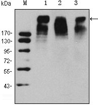
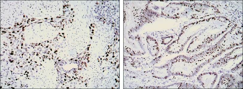
KI67 Antibody
Purified Mouse Monoclonal Antibody
- SPECIFICATION
- CITATIONS
- PROTOCOLS
- BACKGROUND

Application
| WB, IHC, E |
|---|---|
| Primary Accession | P46013 |
| Reactivity | Human |
| Host | Mouse |
| Clonality | Monoclonal |
| Clone Names | 8D5 |
| Isotype | IgG1 |
| Calculated MW | 358kDa |
| Description | Ki67, also known as MKI67, it is the prototypic cell cycle related nuclear protein, expressed by proliferating cells in all phases of the active cell cycle (G1, S, G2 and M phase). It is absent in resting (G0) cells. Ki67 antibodies are useful in establishing the cell growing fraction in neoplasms (immunohistochemically quantified by determining the number of Ki67 positive cells among the total number of resting cells = Ki67 index). In neoplastic tissues the prognostic value is comparable to the tritiated thymidine labelling index. The correlation between low Ki67 index and histologically low grade tumours is strong. Ki67 is routinely used as a neuronal marker of cell cycling and proliferation. |
| Immunogen | Synthetic peptide corresponding to aa (CEDLAGFKELFQTPG) of human KI67, conjugated to KLH. |
| Formulation | Ascitic fluid containing 0.03% sodium azide. |
| Gene ID | 4288 |
|---|---|
| Other Names | Antigen KI-67, MKI67 |
| Dilution | WB~~1/500 - 1/2000 IHC~~1/200 - 1/1000 |
| Storage | Maintain refrigerated at 2-8°C for up to 6 months. For long term storage store at -20°C in small aliquots to prevent freeze-thaw cycles. |
| Precautions | KI67 Antibody is for research use only and not for use in diagnostic or therapeutic procedures. |
| Name | MKI67 (HGNC:7107) |
|---|---|
| Function | Required to maintain individual mitotic chromosomes dispersed in the cytoplasm following nuclear envelope disassembly (PubMed:27362226). Associates with the surface of the mitotic chromosome, the perichromosomal layer, and covers a substantial fraction of the chromosome surface (PubMed:27362226). Prevents chromosomes from collapsing into a single chromatin mass by forming a steric and electrostatic charge barrier: the protein has a high net electrical charge and acts as a surfactant, dispersing chromosomes and enabling independent chromosome motility (PubMed:27362226). Binds DNA, with a preference for supercoiled DNA and AT-rich DNA (PubMed:10878551). Does not contribute to the internal structure of mitotic chromosomes (By similarity). May play a role in chromatin organization (PubMed:24867636). It is however unclear whether it plays a direct role in chromatin organization or whether it is an indirect consequence of its function in maintaining mitotic chromosomes dispersed (Probable). |
| Cellular Location | Chromosome. Nucleus. Nucleus, nucleolus Note=Associates with the surface of the mitotic chromosome, the perichromosomal layer, and covers a substantial fraction of the mitotic chromosome surface (PubMed:27362226). Associates with satellite DNA in G1 phase (PubMed:9510506). Binds tightly to chromatin in interphase, chromatin-binding decreases in mitosis when it associates with the surface of the condensed chromosomes (PubMed:15896774, PubMed:22002106). Predominantly localized in the G1 phase in the perinucleolar region, in the later phases it is also detected throughout the nuclear interior, being predominantly localized in the nuclear matrix (PubMed:22002106). |

Thousands of laboratories across the world have published research that depended on the performance of antibodies from Abcepta to advance their research. Check out links to articles that cite our products in major peer-reviewed journals, organized by research category.
info@abcepta.com, and receive a free "I Love Antibodies" mug.

Provided below are standard protocols that you may find useful for product applications.
References
1. Folia Histochem Cytobiol. 2007;45(4):357-66. 2. Tumori. 2008 May-Jun;94(3):389-97.
If you have used an Abcepta product and would like to share how it has performed, please click on the "Submit Review" button and provide the requested information. Our staff will examine and post your review and contact you if needed.
If you have any additional inquiries please email technical services at tech@abcepta.com.
Ordering Information
Other Products
Shipping Information
