


 Foundational characteristics of cancer include proliferation, angiogenesis, migration, evasion of apoptosis, and cellular immortality. Find key markers for these cellular processes and antibodies to detect them.
 The SUMOplot™ Analysis Program predicts and scores sumoylation sites in your protein. SUMOylation is a post-translational modification involved in various cellular processes, such as nuclear-cytosolic transport, transcriptional regulation, apoptosis, protein stability, response to stress, and progression through the cell cycle.
 The Autophagy Receptor Motif Plotter predicts and scores autophagy receptor binding sites in your protein. Identifying proteins connected to this pathway is critical to understanding the role of autophagy in physiological as well as pathological processes such as development, differentiation, neurodegenerative diseases, stress, infection, and cancer.
BID Antibody
Purified Mouse Monoclonal Antibody


- SPECIFICATION
- CITATIONS
- PROTOCOLS
- BACKGROUND

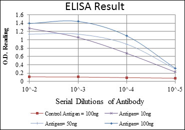
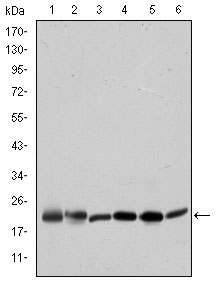
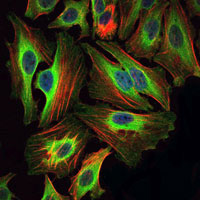
Application
| WB, IHC, FC, ICC, E |
|---|---|
| Primary Accession | P55957 |
| Reactivity | Human |
| Host | Mouse |
| Clonality | Monoclonal |
| Clone Names | 3C5 |
| Isotype | IgG1 |
| Calculated MW | 22kDa |
| Description | This gene encodes a death agonist that heterodimerizes with either agonist BAX or antagonist BCL2. The encoded protein is a member of the BCL-2 family of cell death regulators. It is a mediator of mitochondrial damage induced by caspase-8 (CASP8); CASP8 cleaves this encoded protein, and the COOH-terminal part translocates to mitochondria where it triggers cytochrome c release. Multiple alternatively spliced transcript variants have been found, but the full-length nature of some variants has not been defined.Tissue specificity: Isoform 2 and isoform 3 are expressed in spleen, bone marrow, cerebral and cerebellar cortex. Isoform 2 is expressed in spleen, pancreas and placenta (at protein level). Isoform 3 is expressed in lung, pancreas and spleen (at protein level). Isoform 4 is expressed in lung and pancreas (at protein level) |
| Immunogen | Purified recombinant fragment of human BID expressed in E. Coli. |
| Formulation | Ascitic fluid containing 0.03% sodium azide. |
| Gene ID | 637 |
|---|---|
| Other Names | BH3-interacting domain death agonist, p22 BID, BID, BH3-interacting domain death agonist p15, p15 BID, BH3-interacting domain death agonist p13, p13 BID, BH3-interacting domain death agonist p11, p11 BID, BID |
| Dilution | E~~1/10000 WB~~1/500 - 1/2000 IHC~~1/500 - 1/2000 IF~~1/200 - 1/1000 FC~~1/200 - 1/400 |
| Storage | Maintain refrigerated at 2-8°C for up to 6 months. For long term storage store at -20°C in small aliquots to prevent freeze-thaw cycles. |
| Precautions | BID Antibody is for research use only and not for use in diagnostic or therapeutic procedures. |
| Name | BID |
|---|---|
| Function | Induces caspases and apoptosis (PubMed:14583606). Counters the protective effect of BCL2 (By similarity). |
| Cellular Location | Cytoplasm. Mitochondrion membrane. Mitochondrion outer membrane. Note=When uncleaved, it is predominantly cytoplasmic. [BH3-interacting domain death agonist p13]: Mitochondrion membrane {ECO:0000250|UniProtKB:P70444}. Note=Associated with the mitochondrial membrane. {ECO:0000250|UniProtKB:P70444} [Isoform 3]: Cytoplasm |
| Tissue Location | [Isoform 2]: Expressed in spleen, pancreas and placenta (at protein level). [Isoform 4]: Expressed in lung and pancreas (at protein level). |

Thousands of laboratories across the world have published research that depended on the performance of antibodies from Abcepta to advance their research. Check out links to articles that cite our products in major peer-reviewed journals, organized by research category.
info@abcepta.com, and receive a free "I Love Antibodies" mug.

Provided below are standard protocols that you may find useful for product applications.
References
1. Photochem Photobiol. 2008 Jan-Feb;84(1):250-7. 2. Cell Signal. 2007 Dec;19(12):2468-78.
If you have used an Abcepta product and would like to share how it has performed, please click on the "Submit Review" button and provide the requested information. Our staff will examine and post your review and contact you if needed.
If you have any additional inquiries please email technical services at tech@abcepta.com.
Ordering Information
Other Products
Shipping Information
