


 Foundational characteristics of cancer include proliferation, angiogenesis, migration, evasion of apoptosis, and cellular immortality. Find key markers for these cellular processes and antibodies to detect them.
 The SUMOplot™ Analysis Program predicts and scores sumoylation sites in your protein. SUMOylation is a post-translational modification involved in various cellular processes, such as nuclear-cytosolic transport, transcriptional regulation, apoptosis, protein stability, response to stress, and progression through the cell cycle.
 The Autophagy Receptor Motif Plotter predicts and scores autophagy receptor binding sites in your protein. Identifying proteins connected to this pathway is critical to understanding the role of autophagy in physiological as well as pathological processes such as development, differentiation, neurodegenerative diseases, stress, infection, and cancer.
PRK2 Antibody
Purified Mouse Monoclonal Antibody


- SPECIFICATION
- CITATIONS
- PROTOCOLS
- BACKGROUND

Application
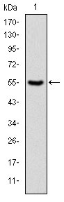
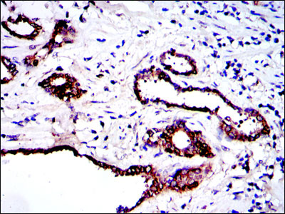
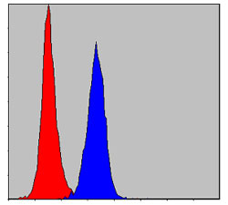
| WB, IHC, FC, E |
|---|---|
| Primary Accession | Q16513 |
| Reactivity | Human, Mouse, Rat, Monkey |
| Host | Mouse |
| Clonality | Monoclonal |
| Clone Names | 1D1 |
| Isotype | IgG1 |
| Calculated MW | 140kDa |
| Description | Protein-kinase-C-related kinases (PRKs) are part of the lipid-regulated protein kinases (PKC) which also include liver PAK & PKN. Human PRK1 and PRK2 share structurally similar catalytic domains, but less similar N-terminal regulatory regions suggesting different regulatory domain functions. PRK1 and PRK2, as well as a third member of this family, PRK3, show distinct patterns of expression in adult tissues. Additionally, the serine-threonine kinase PRK2 can be specifically cleaved by caspase-3 (and/or caspase-3-like subfamily members) during apoptosis. |
| Immunogen | Purified recombinant fragment of human PRK2 expressed in E. Coli. |
| Formulation | Ascitic fluid containing 0.03% sodium azide. |
| Gene ID | 5586 |
|---|---|
| Other Names | Serine/threonine-protein kinase N2, 2.7.11.13, PKN gamma, Protein kinase C-like 2, Protein-kinase C-related kinase 2, PKN2, PRK2, PRKCL2 |
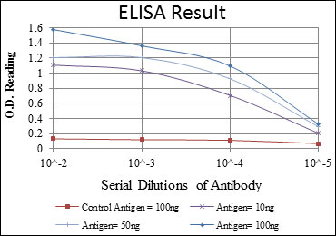
| Dilution | E~~1/10000 WB~~1/500 - 1/2000 IHC~~1/200 - 1/1000 FC~~1/200 - 1/400 |
| Storage | Maintain refrigerated at 2-8°C for up to 6 months. For long term storage store at -20°C in small aliquots to prevent freeze-thaw cycles. |
| Precautions | PRK2 Antibody is for research use only and not for use in diagnostic or therapeutic procedures. |
| Name | PKN2 |
|---|---|
| Synonyms | PRK2, PRKCL2 |
| Function | PKC-related serine/threonine-protein kinase and Rho/Rac effector protein that participates in specific signal transduction responses in the cell. Plays a role in the regulation of cell cycle progression, actin cytoskeleton assembly, cell migration, cell adhesion, tumor cell invasion and transcription activation signaling processes. Phosphorylates CTTN in hyaluronan-induced astrocytes and hence decreases CTTN ability to associate with filamentous actin. Phosphorylates HDAC5, therefore lead to impair HDAC5 import. Direct RhoA target required for the regulation of the maturation of primordial junctions into apical junction formation in bronchial epithelial cells. Required for G2/M phases of the cell cycle progression and abscission during cytokinesis in a ECT2-dependent manner. Stimulates FYN kinase activity that is required for establishment of skin cell-cell adhesion during keratinocytes differentiation. Regulates epithelial bladder cells speed and direction of movement during cell migration and tumor cell invasion. Inhibits Akt pro-survival-induced kinase activity. Mediates Rho protein-induced transcriptional activation via the c-fos serum response factor (SRF). Involved in the negative regulation of ciliogenesis (PubMed:27104747). |
| Cellular Location | Cytoplasm. Nucleus Membrane {ECO:0000250|UniProtKB:Q8BWW9}. Cell projection, lamellipodium. Cytoplasm, cytoskeleton. Cleavage furrow. Midbody Cell junction. Note=Colocalizes with PTPN13 in lamellipodia-like structures, regions of large actin turnover. Accumulates during telophase at the cleavage furrow and concentrates finally around the midbody in cytokinesis. Recruited to nascent cell-cell contacts at the apical surface of cells. In the course of viral infection, colocalizes with HCV NS5B at perinuclear region in the cytoplasm. |
| Tissue Location | Ubiquitous. Expressed in numerous tumor cell lines, especially in bladder tumor cells. |

Thousands of laboratories across the world have published research that depended on the performance of antibodies from Abcepta to advance their research. Check out links to articles that cite our products in major peer-reviewed journals, organized by research category.
info@abcepta.com, and receive a free "I Love Antibodies" mug.

Provided below are standard protocols that you may find useful for product applications.
References
1. Cell. 2009 Jul 23;138(2):389-403. 2. Ann Intern Med. 2009 Apr 21;150(8):541-50.
If you have used an Abcepta product and would like to share how it has performed, please click on the "Submit Review" button and provide the requested information. Our staff will examine and post your review and contact you if needed.
If you have any additional inquiries please email technical services at tech@abcepta.com.
Ordering Information
Other Products
Shipping Information
