


 Foundational characteristics of cancer include proliferation, angiogenesis, migration, evasion of apoptosis, and cellular immortality. Find key markers for these cellular processes and antibodies to detect them.
 The SUMOplot™ Analysis Program predicts and scores sumoylation sites in your protein. SUMOylation is a post-translational modification involved in various cellular processes, such as nuclear-cytosolic transport, transcriptional regulation, apoptosis, protein stability, response to stress, and progression through the cell cycle.
 The Autophagy Receptor Motif Plotter predicts and scores autophagy receptor binding sites in your protein. Identifying proteins connected to this pathway is critical to understanding the role of autophagy in physiological as well as pathological processes such as development, differentiation, neurodegenerative diseases, stress, infection, and cancer.
CDKN2A Antibody
Purified Mouse Monoclonal Antibody


- SPECIFICATION
- CITATIONS
- PROTOCOLS
- BACKGROUND

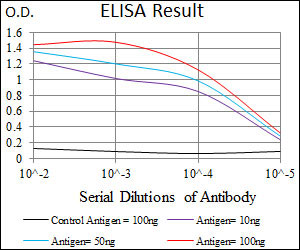
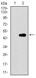
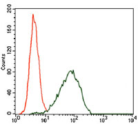
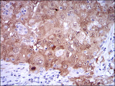
Application
| WB, IHC, FC, E |
|---|---|
| Primary Accession | P42771 |
| Reactivity | Human |
| Host | Mouse |
| Clonality | Monoclonal |
| Clone Names | 1D7D2 |
| Isotype | IgG1 |
| Calculated MW | 16.5kDa |
| Description | This gene generates several transcript variants which differ in their first exons. At least three alternatively spliced variants encoding distinct proteins have been reported, two of which encode structurally related isoforms known to function as inhibitors of CDK4 kinase. The remaining transcript includes an alternate first exon located 20 Kb upstream of the remainder of the gene; this transcript contains an alternate open reading frame (ARF) that specifies a protein which is structurally unrelated to the products of the other variants. This ARF product functions as a stabilizer of the tumor suppressor protein p53 as it can interact with, and sequester, MDM1, a protein responsible for the degradation of p53. In spite of the structural and functional differences, the CDK inhibitor isoforms and the ARF product encoded by this gene, through the regulatory roles of CDK4 and p53 in cell cycle G1 progression, share a common functionality in cell cycle G1 control. This gene is frequently mutated or deleted in a wide variety of tumors, and is known to be an important tumor suppressor gene. |
| Immunogen | Purified recombinant fragment of human CDKN2A (AA: 1-156) expressed in E. Coli. |
| Formulation | Ascitic fluid containing 0.03% sodium azide. |
| Gene ID | 1029 |
|---|---|
| Other Names | Cyclin-dependent kinase inhibitor 2A, isoforms 1/2/3, Cyclin-dependent kinase 4 inhibitor A, CDK4I, Multiple tumor suppressor 1, MTS-1, p16-INK4a, p16-INK4, p16INK4A, CDKN2A, CDKN2, MTS1 |
| Dilution | E~~1/10000 WB~~1/500 - 1/2000 FC~~1/200 - 1/400 IHC~~1/200 - 1/1000 |
| Storage | Maintain refrigerated at 2-8°C for up to 6 months. For long term storage store at -20°C in small aliquots to prevent freeze-thaw cycles. |
| Precautions | CDKN2A Antibody is for research use only and not for use in diagnostic or therapeutic procedures. |
| Name | CDKN2A (HGNC:1787) |
|---|---|
| Synonyms | CDKN2, MTS1 |
| Function | Acts as a negative regulator of the proliferation of normal cells by interacting strongly with CDK4 and CDK6. This inhibits their ability to interact with cyclins D and to phosphorylate the retinoblastoma protein. |
| Cellular Location | Cytoplasm. Nucleus |
| Tissue Location | Widely expressed but not detected in brain or skeletal muscle. Isoform 3 is pancreas-specific |

Thousands of laboratories across the world have published research that depended on the performance of antibodies from Abcepta to advance their research. Check out links to articles that cite our products in major peer-reviewed journals, organized by research category.
info@abcepta.com, and receive a free "I Love Antibodies" mug.

Provided below are standard protocols that you may find useful for product applications.
References
1.Clin Cancer Res. 2011 Dec 1;17(23):7413-23. 2.Appl Immunohistochem Mol Morphol. 2011 Dec;19(6):562-8.
If you have used an Abcepta product and would like to share how it has performed, please click on the "Submit Review" button and provide the requested information. Our staff will examine and post your review and contact you if needed.
If you have any additional inquiries please email technical services at tech@abcepta.com.
Ordering Information
Other Products
Shipping Information
