PKN1 Antibody
Purified Mouse Monoclonal Antibody
- SPECIFICATION
- CITATIONS
- PROTOCOLS
- BACKGROUND

Application
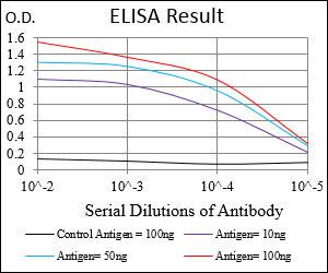
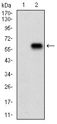
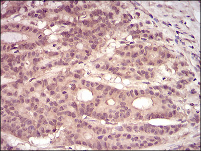
| WB, IHC, FC, ICC, E |
|---|---|
| Primary Accession | Q16512 |
| Reactivity | Human |
| Host | Mouse |
| Clonality | Monoclonal |
| Clone Names | 4H10B1 |
| Isotype | IgG2b |
| Calculated MW | 104kDa |
| Description | The protein encoded by this gene belongs to the protein kinase C superfamily. This kinase is activated by Rho family of small G proteins and may mediate the Rho-dependent signaling pathway. This kinase can be activated by phospholipids and by limited proteolysis. The 3-phosphoinositide dependent protein kinase-1 (PDPK1/PDK1) is reported to phosphorylate this kinase, which may mediate insulin signals to the actin cytoskeleton. The proteolytic activation of this kinase by caspase-3 or related proteases during apoptosis suggests its role in signal transduction related to apoptosis. Alternatively spliced transcript variants encoding distinct isoforms have been observed. |
| Immunogen | Purified recombinant fragment of human PKN1 (AA: 442-620) expressed in E. Coli. |
| Formulation | Purified antibody in PBS with 0.05% sodium azide. |
| Gene ID | 5585 |
|---|---|
| Other Names | Serine/threonine-protein kinase N1, 2.7.11.13, Protease-activated kinase 1, PAK-1, Protein kinase C-like 1, Protein kinase C-like PKN, Protein kinase PKN-alpha, Protein-kinase C-related kinase 1, Serine-threonine protein kinase N, PKN1, PAK1, PKN, PRK1, PRKCL1 |
| Dilution | E~~1/10000 WB~~1/500 - 1/2000 IF~~1/200 - 1/1000 FC~~1/200 - 1/400 IHC~~1/200 - 1/1000 |
| Storage | Maintain refrigerated at 2-8°C for up to 6 months. For long term storage store at -20°C in small aliquots to prevent freeze-thaw cycles. |
| Precautions | PKN1 Antibody is for research use only and not for use in diagnostic or therapeutic procedures. |
| Name | PKN1 |
|---|---|
| Synonyms | PAK1, PKN, PRK1, PRKCL1 |
| Function | PKC-related serine/threonine-protein kinase involved in various processes such as regulation of the intermediate filaments of the actin cytoskeleton, cell migration, tumor cell invasion and transcription regulation. Part of a signaling cascade that begins with the activation of the adrenergic receptor ADRA1B and leads to the activation of MAPK14. Regulates the cytoskeletal network by phosphorylating proteins such as VIM and neurofilament proteins NEFH, NEFL and NEFM, leading to inhibit their polymerization. Phosphorylates 'Ser-575', 'Ser-637' and 'Ser-669' of MAPT/Tau, lowering its ability to bind to microtubules, resulting in disruption of tubulin assembly. Acts as a key coactivator of androgen receptor (AR)-dependent transcription, by being recruited to AR target genes and specifically mediating phosphorylation of 'Thr-11' of histone H3 (H3T11ph), a specific tag for epigenetic transcriptional activation that promotes demethylation of histone H3 'Lys-9' (H3K9me) by KDM4C/JMJD2C. Phosphorylates HDAC5, HDAC7 and HDAC9, leading to impair their import in the nucleus. Phosphorylates 'Thr-38' of PPP1R14A, 'Ser-159', 'Ser-163' and 'Ser-170' of MARCKS, and GFAP. Able to phosphorylate RPS6 in vitro. |
| Cellular Location | Cytoplasm. Nucleus Endosome. Cell membrane {ECO:0000250|UniProtKB:Q63433}; Peripheral membrane protein {ECO:0000250|UniProtKB:Q63433}. Cleavage furrow. Midbody Note=Associates with chromatin in a ligand-dependent manner Localization to endosomes is mediated via its interaction with RHOB Association to the cell membrane is dependent on Ser-377 phosphorylation. Accumulates during telophase at the cleavage furrow and finally concentrates around the midbody in cytokinesis {ECO:0000250|UniProtKB:Q63433, ECO:0000269|PubMed:17332740} |
| Tissue Location | Found ubiquitously. Expressed in heart, brain, placenta, lung, skeletal muscle, kidney and pancreas. Expressed in numerous tumor cell lines, especially in breast tumor cells |

Thousands of laboratories across the world have published research that depended on the performance of antibodies from Abcepta to advance their research. Check out links to articles that cite our products in major peer-reviewed journals, organized by research category.
info@abcepta.com, and receive a free "I Love Antibodies" mug.
Provided below are standard protocols that you may find useful for product applications.
Background
T protein p53 binding protein 1 may have a role in checkpoint signaling during mitosis,enhance TP53-mediated transcriptional activation and play a role in the response to DNA damage. ;
References
1. J Biol Chem. 2013 Nov 29;288(48):34658-70.2. Hum Pathol. 2009 Oct;40(10):1434-40.
If you have used an Abcepta product and would like to share how it has performed, please click on the "Submit Review" button and provide the requested information. Our staff will examine and post your review and contact you if needed.
If you have any additional inquiries please email technical services at tech@abcepta.com.


 Foundational characteristics of cancer include proliferation, angiogenesis, migration, evasion of apoptosis, and cellular immortality. Find key markers for these cellular processes and antibodies to detect them.
 The SUMOplot™ Analysis Program predicts and scores sumoylation sites in your protein. SUMOylation is a post-translational modification involved in various cellular processes, such as nuclear-cytosolic transport, transcriptional regulation, apoptosis, protein stability, response to stress, and progression through the cell cycle.
 The Autophagy Receptor Motif Plotter predicts and scores autophagy receptor binding sites in your protein. Identifying proteins connected to this pathway is critical to understanding the role of autophagy in physiological as well as pathological processes such as development, differentiation, neurodegenerative diseases, stress, infection, and cancer.


