


 Foundational characteristics of cancer include proliferation, angiogenesis, migration, evasion of apoptosis, and cellular immortality. Find key markers for these cellular processes and antibodies to detect them.
 The SUMOplot™ Analysis Program predicts and scores sumoylation sites in your protein. SUMOylation is a post-translational modification involved in various cellular processes, such as nuclear-cytosolic transport, transcriptional regulation, apoptosis, protein stability, response to stress, and progression through the cell cycle.
 The Autophagy Receptor Motif Plotter predicts and scores autophagy receptor binding sites in your protein. Identifying proteins connected to this pathway is critical to understanding the role of autophagy in physiological as well as pathological processes such as development, differentiation, neurodegenerative diseases, stress, infection, and cancer.
Rab13 Antibody
Purified Mouse Monoclonal Antibody

- SPECIFICATION
- CITATIONS
- PROTOCOLS
- BACKGROUND

Application
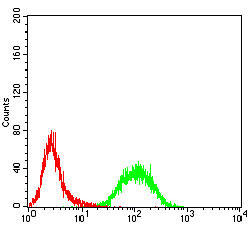
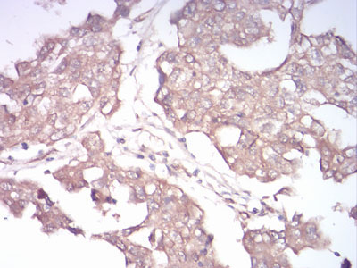
| WB, IHC, FC, ICC, E |
|---|---|
| Primary Accession | P51153 |
| Reactivity | Human |
| Host | Mouse |
| Clonality | Monoclonal |
| Clone Names | 7G8A4 |
| Isotype | IgG1 |
| Calculated MW | 22.8kDa |
| Description | This gene is a member of the Rab family of small G proteins and plays a role in regulating membrane trafficking between trans-Golgi network (TGN) and recycling endosomes (RE). The encoded protein is involved in the assembly of tight junctions, which are components of the apical junctional complex (AJC) of epithelial cells. The AJC plays a role in forming a barrier between luminal contents and the underlying tissue. Additional functions associated with the protein include endocytic recycling of occludin, regulation of epithelial cell scattering, neuronal regeneration and regulation of neurite outgrowth. Alternately spliced transcript variants have been observed for this gene. A pseudogene associated with this gene is located on chromosome 12. |
| Immunogen | Purified recombinant fragment of human Rab13 (AA: 66-200) expressed in E. Coli. |
| Formulation | Purified antibody in PBS with 0.05% sodium azide |
| Gene ID | 5872 |
|---|---|
| Other Names | Ras-related protein Rab-13, Cell growth-inhibiting gene 4 protein, RAB13 |
| Dilution | E~~1/10000 WB~~1/500 - 1/2000 IF~~1/200 - 1/1000 FC~~1/200 - 1/400 IHC~~1/200 - 1/1000 |
| Storage | Maintain refrigerated at 2-8°C for up to 6 months. For long term storage store at -20°C in small aliquots to prevent freeze-thaw cycles. |
| Precautions | Rab13 Antibody is for research use only and not for use in diagnostic or therapeutic procedures. |
| Name | RAB13 |
|---|---|
| Function | The small GTPases Rab are key regulators of intracellular membrane trafficking, from the formation of transport vesicles to their fusion with membranes. Rabs cycle between an inactive GDP-bound form and an active GTP-bound form that is able to recruit to membranes different sets of downstream effectors directly responsible for vesicle formation, movement, tethering and fusion. That Rab is involved in endocytic recycling and regulates the transport to the plasma membrane of transmembrane proteins like the tight junction protein OCLN/occludin. Thereby, it regulates the assembly and the activity of tight junctions. Moreover, it may also regulate tight junction assembly by activating the PKA signaling pathway and by reorganizing the actin cytoskeleton through the activation of the downstream effectors PRKACA and MICALL2 respectively. Through its role in tight junction assembly, may play a role in the establishment of Sertoli cell barrier. Plays also a role in angiogenesis through regulation of endothelial cells chemotaxis. Also involved in neurite outgrowth. Has also been proposed to play a role in post-Golgi membrane trafficking from the TGN to the recycling endosome. Finally, it has been involved in insulin-induced transport to the plasma membrane of the glucose transporter GLUT4 and therefore may play a role in glucose homeostasis. |
| Cellular Location | Cell membrane; Lipid-anchor; Cytoplasmic side. Cytoplasmic vesicle membrane; Lipid-anchor; Cytoplasmic side. Cell junction, tight junction. Golgi apparatus, trans-Golgi network membrane Recycling endosome membrane. Cell projection, lamellipodium {ECO:0000250|UniProtKB:Q9DD03}. Note=Tight junctions or associated with vesicles scattered throughout the cytoplasm in cells lacking tight junctions (PubMed:8294494) Relocalizes to the leading edge of lamellipodia in migrating endothelial cells (By similarity). {ECO:0000250|UniProtKB:Q9DD03, ECO:0000269|PubMed:8294494} |
| Tissue Location | Detected in several types of epithelia, including intestine, kidney, liver and in endothelial cells |

Thousands of laboratories across the world have published research that depended on the performance of antibodies from Abcepta to advance their research. Check out links to articles that cite our products in major peer-reviewed journals, organized by research category.
info@abcepta.com, and receive a free "I Love Antibodies" mug.

Provided below are standard protocols that you may find useful for product applications.
References
1.J Histochem Cytochem. 2012 Jul;60(7):537-49.2.J Cell Biol. 2008 Sep 8;182(5):845-53.
If you have used an Abcepta product and would like to share how it has performed, please click on the "Submit Review" button and provide the requested information. Our staff will examine and post your review and contact you if needed.
If you have any additional inquiries please email technical services at tech@abcepta.com.
Ordering Information
Other Products
Shipping Information
