


 Foundational characteristics of cancer include proliferation, angiogenesis, migration, evasion of apoptosis, and cellular immortality. Find key markers for these cellular processes and antibodies to detect them.
 The SUMOplot™ Analysis Program predicts and scores sumoylation sites in your protein. SUMOylation is a post-translational modification involved in various cellular processes, such as nuclear-cytosolic transport, transcriptional regulation, apoptosis, protein stability, response to stress, and progression through the cell cycle.
 The Autophagy Receptor Motif Plotter predicts and scores autophagy receptor binding sites in your protein. Identifying proteins connected to this pathway is critical to understanding the role of autophagy in physiological as well as pathological processes such as development, differentiation, neurodegenerative diseases, stress, infection, and cancer.
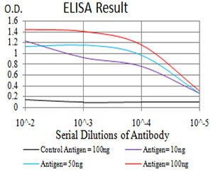
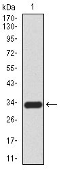
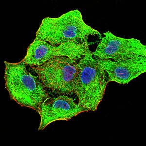
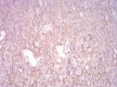
CK5 Antibody
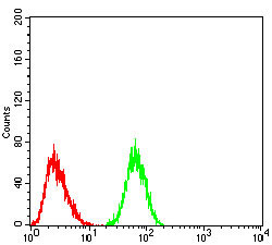
Purified Mouse Monoclonal Antibody


- SPECIFICATION
- CITATIONS
- PROTOCOLS
- BACKGROUND

Application
| WB, IHC, FC, ICC, E |
|---|---|
| Primary Accession | P13647 |
| Reactivity | Human, Mouse, Monkey |
| Host | Mouse |
| Clonality | Monoclonal |
| Clone Names | 3D6D4 |
| Isotype | IgG1 |
| Calculated MW | 62.4kDa |
| Description | The protein encoded by this gene is a member of the keratin gene family. The type II cytokeratins consist of basic or neutral proteins which are arranged in pairs of heterotypic keratin chains coexpressed during differentiation of simple and stratified epithelial tissues. This type II cytokeratin is specifically expressed in the basal layer of the epidermis with family member KRT14. Mutations in these genes have been associated with a complex of diseases termed epidermolysis bullosa simplex. The type II cytokeratins are clustered in a region of chromosome 12q12-q13. |
| Immunogen | Purified recombinant fragment of human CK5 (AA: 258-357) expressed in E. Coli. |
| Formulation | Purified antibody in PBS with 0.05% sodium azide |
| Gene ID | 3852 |
|---|---|
| Other Names | Keratin, type II cytoskeletal 5, 58 kDa cytokeratin, Cytokeratin-5, CK-5, Keratin-5, K5, Type-II keratin Kb5, KRT5 |
| Dilution | E~~1/10000 WB~~1/500 - 1/2000 IF~~1/200 - 1/1000 IHC~~1/200 - 1/1000 FC~~1/200 - 1/400 |
| Storage | Maintain refrigerated at 2-8°C for up to 6 months. For long term storage store at -20°C in small aliquots to prevent freeze-thaw cycles. |
| Precautions | CK5 Antibody is for research use only and not for use in diagnostic or therapeutic procedures. |
| Name | KRT5 |
|---|---|
| Function | Required for the formation of keratin intermediate filaments in the basal epidermis and maintenance of the skin barrier in response to mechanical stress (By similarity). Regulates the recruitment of Langerhans cells to the epidermis, potentially by modulation of the abundance of macrophage chemotactic cytokines, macrophage inflammatory cytokines and CTNND1 localization in keratinocytes (By similarity). |
| Cellular Location | Cytoplasm. |
| Tissue Location | Expressed in corneal epithelium (at protein level) (PubMed:26758872). Expressed in keratinocytes (at protein level) (PubMed:20128788, PubMed:31302245). |

Thousands of laboratories across the world have published research that depended on the performance of antibodies from Abcepta to advance their research. Check out links to articles that cite our products in major peer-reviewed journals, organized by research category.
info@abcepta.com, and receive a free "I Love Antibodies" mug.

Provided below are standard protocols that you may find useful for product applications.
References
1.Horm Cancer. 2013 Feb;4(1):36-49.2.Breast Cancer Res Treat. 2011 Jul;128(1):45-55.
If you have used an Abcepta product and would like to share how it has performed, please click on the "Submit Review" button and provide the requested information. Our staff will examine and post your review and contact you if needed.
If you have any additional inquiries please email technical services at tech@abcepta.com.
Ordering Information
Other Products
Shipping Information
