


 Foundational characteristics of cancer include proliferation, angiogenesis, migration, evasion of apoptosis, and cellular immortality. Find key markers for these cellular processes and antibodies to detect them.
 The SUMOplot™ Analysis Program predicts and scores sumoylation sites in your protein. SUMOylation is a post-translational modification involved in various cellular processes, such as nuclear-cytosolic transport, transcriptional regulation, apoptosis, protein stability, response to stress, and progression through the cell cycle.
 The Autophagy Receptor Motif Plotter predicts and scores autophagy receptor binding sites in your protein. Identifying proteins connected to this pathway is critical to understanding the role of autophagy in physiological as well as pathological processes such as development, differentiation, neurodegenerative diseases, stress, infection, and cancer.
Mouse Monoclonal Antibody to ALK
Purified Mouse Monoclonal Antibody
- SPECIFICATION
- CITATIONS
- PROTOCOLS
- BACKGROUND

Application
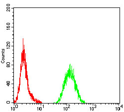
| WB, FC, ICC, E |
|---|---|
| Primary Accession | Q9UM73 |
| Reactivity | Human |
| Host | Mouse |
| Clonality | Monoclonal |
| Clone Names | 7A11A4 |
| Isotype | Mouse IgG1 |
| Calculated MW | 176kDa |
| Description | This gene encodes a receptor tyrosine kinase, which belongs to the insulin receptor superfamily. This protein comprises an extracellular domain, an hydrophobic stretch corresponding to a single pass transmembrane region, and an intracellular kinase domain. It plays an important role in the development of the brain and exerts its effects on specific neurons in the nervous system. This gene has been found to be rearranged, mutated, or amplified in a series of tumours including anaplastic large cell lymphomas, neuroblastoma, and non-small cell lung cancer. The chromosomal rearrangements are the most common genetic alterations in this gene, which result in creation of multiple fusion genes in tumourigenesis, including ALK (chromosome 2)/EML4 (chromosome 2), ALK/RANBP2 (chromosome 2), ALK/ATIC (chromosome 2), ALK/TFG (chromosome 3), ALK/NPM1 (chromosome 5), ALK/SQSTM1 (chromosome 5), ALK/KIF5B (chromosome 10), ALK/CLTC (chromosome 17), ALK/TPM4 (chromosome 19), and ALK/MSN (chromosome X).; |
| Immunogen | Purified recombinant fragment of human ALK (AA: 1366-1468) expressed in E. Coli. |
| Formulation | Purified antibody in PBS with 0.05% sodium azide |
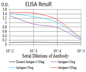
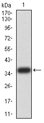
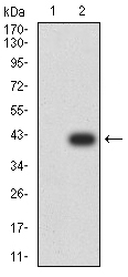
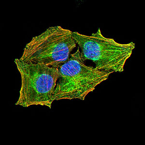
| Application Note | ELISA: 1/10000 WB: 1/500 - 1/2000 ICC: 1/50 - 1/250 FCM: 1/200 - 1/400 |
| Gene ID | 238 |
|---|---|
| Other Names | CD246; NBLST3 |
| Storage | Maintain refrigerated at 2-8°C for up to 6 months. For long term storage store at -20°C in small aliquots to prevent freeze-thaw cycles. |
| Precautions | Mouse Monoclonal Antibody to ALK is for research use only and not for use in diagnostic or therapeutic procedures. |
| Name | ALK {ECO:0000303|PubMed:9174053, ECO:0000312|HGNC:HGNC:427} |
|---|---|
| Function | Neuronal receptor tyrosine kinase that is essentially and transiently expressed in specific regions of the central and peripheral nervous systems and plays an important role in the genesis and differentiation of the nervous system (PubMed:11121404, PubMed:11387242, PubMed:16317043, PubMed:17274988, PubMed:30061385, PubMed:34646012, PubMed:34819673). Also acts as a key thinness protein involved in the resistance to weight gain: in hypothalamic neurons, controls energy expenditure acting as a negative regulator of white adipose tissue lipolysis and sympathetic tone to fine-tune energy homeostasis (By similarity). Following activation by ALKAL2 ligand at the cell surface, transduces an extracellular signal into an intracellular response (PubMed:30061385, PubMed:33411331, PubMed:34646012, PubMed:34819673). In contrast, ALKAL1 is not a potent physiological ligand for ALK (PubMed:34646012). Ligand-binding to the extracellular domain induces tyrosine kinase activation, leading to activation of the mitogen-activated protein kinase (MAPK) pathway (PubMed:34819673). Phosphorylates almost exclusively at the first tyrosine of the Y-x-x-x-Y-Y motif (PubMed:15226403, PubMed:16878150). Induces tyrosine phosphorylation of CBL, FRS2, IRS1 and SHC1, as well as of the MAP kinases MAPK1/ERK2 and MAPK3/ERK1 (PubMed:15226403, PubMed:16878150). ALK activation may also be regulated by pleiotrophin (PTN) and midkine (MDK) (PubMed:11278720, PubMed:11809760, PubMed:12107166, PubMed:12122009). PTN-binding induces MAPK pathway activation, which is important for the anti-apoptotic signaling of PTN and regulation of cell proliferation (PubMed:11278720, PubMed:11809760, PubMed:12107166). MDK-binding induces phosphorylation of the ALK target insulin receptor substrate (IRS1), activates mitogen-activated protein kinases (MAPKs) and PI3-kinase, resulting also in cell proliferation induction (PubMed:12122009). Drives NF-kappa-B activation, probably through IRS1 and the activation of the AKT serine/threonine kinase (PubMed:15226403, PubMed:16878150). Recruitment of IRS1 to activated ALK and the activation of NF-kappa-B are essential for the autocrine growth and survival signaling of MDK (PubMed:15226403, PubMed:16878150). |
| Cellular Location | Cell membrane; Single-pass type I membrane protein Note=Membrane attachment is essential for promotion of neuron-like differentiation and cell proliferation arrest through specific activation of the MAP kinase pathway. |
| Tissue Location | Expressed in brain and CNS. Also expressed in the small intestine and testis, but not in normal lymphoid cells |

Thousands of laboratories across the world have published research that depended on the performance of antibodies from Abcepta to advance their research. Check out links to articles that cite our products in major peer-reviewed journals, organized by research category.
info@abcepta.com, and receive a free "I Love Antibodies" mug.

Provided below are standard protocols that you may find useful for product applications.
References
1.Nature. 2015 Oct 15;526(7573):453-7. ; 2.Breast Cancer Res. 2015 Sep 17;17:127.;
If you have used an Abcepta product and would like to share how it has performed, please click on the "Submit Review" button and provide the requested information. Our staff will examine and post your review and contact you if needed.
If you have any additional inquiries please email technical services at tech@abcepta.com.
Ordering Information
Other Products
Shipping Information
