


 Foundational characteristics of cancer include proliferation, angiogenesis, migration, evasion of apoptosis, and cellular immortality. Find key markers for these cellular processes and antibodies to detect them.
 The SUMOplot™ Analysis Program predicts and scores sumoylation sites in your protein. SUMOylation is a post-translational modification involved in various cellular processes, such as nuclear-cytosolic transport, transcriptional regulation, apoptosis, protein stability, response to stress, and progression through the cell cycle.
 The Autophagy Receptor Motif Plotter predicts and scores autophagy receptor binding sites in your protein. Identifying proteins connected to this pathway is critical to understanding the role of autophagy in physiological as well as pathological processes such as development, differentiation, neurodegenerative diseases, stress, infection, and cancer.
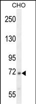
C2 Antibody (N-term)
Affinity Purified Rabbit Polyclonal Antibody (Pab)
- SPECIFICATION
- CITATIONS
- PROTOCOLS
- BACKGROUND

Application
| WB, E |
|---|---|
| Primary Accession | P06681 |
| Other Accession | NP_000054 |
| Reactivity | Human, Hamster |
| Host | Rabbit |
| Clonality | Polyclonal |
| Isotype | Rabbit IgG |
| Calculated MW | 83268 Da |
| Antigen Region | 147-176 aa |
| Gene ID | 717 |
|---|---|
| Other Names | Complement C2, C3/C5 convertase, Complement C2b fragment, Complement C2a fragment, C2 |
| Target/Specificity | This C2 antibody is generated from rabbits immunized with a KLH conjugated synthetic peptide between 147-176 amino acids from the N-terminal region of human C2. |
| Dilution | WB~~1:1000 |
| Format | Purified polyclonal antibody supplied in PBS with 0.09% (W/V) sodium azide. This antibody is purified through a protein A column, followed by peptide affinity purification. |
| Storage | Maintain refrigerated at 2-8°C for up to 2 weeks. For long term storage store at -20°C in small aliquots to prevent freeze-thaw cycles. |
| Precautions | C2 Antibody (N-term) is for research use only and not for use in diagnostic or therapeutic procedures. |
| Name | C2 |
|---|---|
| Function | Component C2 which is part of the classical pathway of the complement system is cleaved by activated factor C1 into two fragments: C2b and C2a. C2a, a serine protease, then combines with complement factor C4b to generate the C3 or C5 convertase. |
| Cellular Location | Secreted. |

Thousands of laboratories across the world have published research that depended on the performance of antibodies from Abcepta to advance their research. Check out links to articles that cite our products in major peer-reviewed journals, organized by research category.
info@abcepta.com, and receive a free "I Love Antibodies" mug.

Provided below are standard protocols that you may find useful for product applications.
Background
Component C2 is a serum glycoprotein that functions as part of the classical pathway of the complement system. Activated C1 cleaves C2 into C2a and C2b. The serine proteinase C2a then combines with complement factor 4b to create the C3 or C5 convertase. Deficiency of C2 has been reported to associated with certain autoimmune diseases and SNPs in this gene have been associated with altered susceptibility to age-related macular degeneration. This gene localizes within the class III region of the MHC on the short arm of chromosome 6. Alternative splicing results in multiple transcript variants encoding distinct isoforms. Additional transcript variants have been described in publications but their full-length sequence has not been determined.
References
Hu, M., et al. Pharmacogenet. Genomics 20(10):634-637(2010)
Bailey, S.D., et al. Diabetes Care 33(10):2250-2253(2010)
Liu, X., et al. Retina (Philadelphia, Pa.) 30(8):1177-1184(2010)
Han, S., et al. Hum. Immunol. 71(7):727-730(2010)
Ishii, Y., et al. J. Immunol. 151(1):170-174(1993)
If you have used an Abcepta product and would like to share how it has performed, please click on the "Submit Review" button and provide the requested information. Our staff will examine and post your review and contact you if needed.
If you have any additional inquiries please email technical services at tech@abcepta.com.
Ordering Information
Other Products
Shipping Information
