


 Foundational characteristics of cancer include proliferation, angiogenesis, migration, evasion of apoptosis, and cellular immortality. Find key markers for these cellular processes and antibodies to detect them.
 The SUMOplot™ Analysis Program predicts and scores sumoylation sites in your protein. SUMOylation is a post-translational modification involved in various cellular processes, such as nuclear-cytosolic transport, transcriptional regulation, apoptosis, protein stability, response to stress, and progression through the cell cycle.
 The Autophagy Receptor Motif Plotter predicts and scores autophagy receptor binding sites in your protein. Identifying proteins connected to this pathway is critical to understanding the role of autophagy in physiological as well as pathological processes such as development, differentiation, neurodegenerative diseases, stress, infection, and cancer.
RORA Antibody (T216)
Affinity Purified Rabbit Polyclonal Antibody (Pab)

- SPECIFICATION
- CITATIONS
- PROTOCOLS
- BACKGROUND

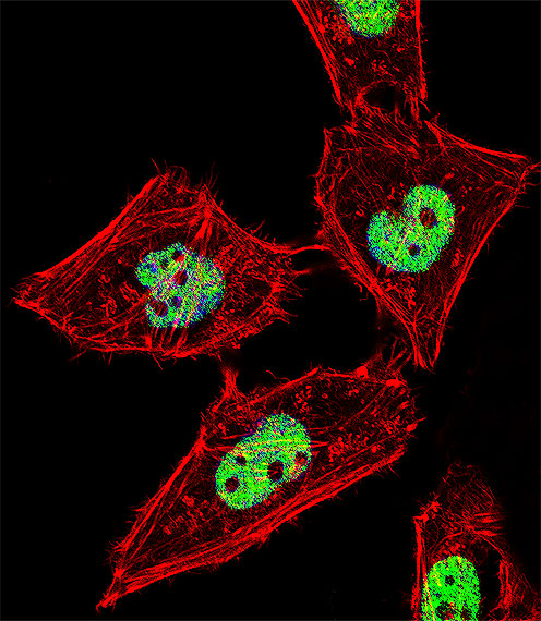
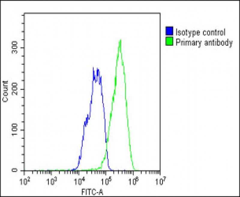
Application
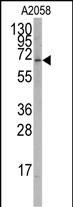
| WB, IF, FC, E |
|---|---|
| Primary Accession | P35398 |
| Other Accession | P51448, NP_599022 |
| Reactivity | Human |
| Predicted | Mouse |
| Host | Rabbit |
| Clonality | Polyclonal |
| Isotype | Rabbit IgG |
| Calculated MW | 58975 Da |
| Antigen Region | 193-222 aa |
| Gene ID | 6095 |
|---|---|
| Other Names | Nuclear receptor ROR-alpha, Nuclear receptor RZR-alpha, Nuclear receptor subfamily 1 group F member 1, RAR-related orphan receptor A, Retinoid-related orphan receptor-alpha, RORA, NR1F1, RZRA |
| Target/Specificity | This RORA antibody is generated from rabbits immunized with a KLH conjugated synthetic peptide between 193-222 amino acids from human RORA. |
| Dilution | IF~~1:10~50 WB~~1:500 FC~~1:25 |
| Format | Purified polyclonal antibody supplied in PBS with 0.09% (W/V) sodium azide. This antibody is purified through a protein A column, followed by peptide affinity purification. |
| Storage | Maintain refrigerated at 2-8°C for up to 2 weeks. For long term storage store at -20°C in small aliquots to prevent freeze-thaw cycles. |
| Precautions | RORA Antibody (T216) is for research use only and not for use in diagnostic or therapeutic procedures. |
| Name | RORA |
|---|---|
| Synonyms | NR1F1, RZRA |
| Function | Nuclear receptor that binds DNA as a monomer to ROR response elements (RORE) containing a single core motif half-site 5'-AGGTCA-3' preceded by a short A-T-rich sequence. Key regulator of embryonic development, cellular differentiation, immunity, circadian rhythm as well as lipid, steroid, xenobiotics and glucose metabolism. Considered to have intrinsic transcriptional activity, have some natural ligands like oxysterols that act as agonists (25-hydroxycholesterol) or inverse agonists (7-oxygenated sterols), enhancing or repressing the transcriptional activity, respectively. Recruits distinct combinations of cofactors to target genes regulatory regions to modulate their transcriptional expression, depending on the tissue, time and promoter contexts. Regulates genes involved in photoreceptor development including OPN1SW, OPN1SM and ARR3 and skeletal muscle development with MYOD1. Required for proper cerebellum development (PubMed:29656859). Regulates SHH gene expression, among others, to induce granule cells proliferation as well as expression of genes involved in calcium- mediated signal transduction. Regulates the circadian expression of several clock genes, including CLOCK, BMAL1, NPAS2 and CRY1. Competes with NR1D1 for binding to their shared DNA response element on some clock genes such as BMAL1, CRY1 and NR1D1 itself, resulting in NR1D1- mediated repression or RORA-mediated activation of clock genes expression, leading to the circadian pattern of clock genes expression. Therefore influences the period length and stability of the clock. Regulates genes involved in lipid metabolism such as apolipoproteins APOA1, APOA5, APOC3 and PPARG. In liver, has specific and redundant functions with RORC as positive or negative modulator of expression of genes encoding phase I and phase II proteins involved in the metabolism of lipids, steroids and xenobiotics, such as CYP7B1 and SULT2A1. Induces a rhythmic expression of some of these genes. In addition, interplays functionally with NR1H2 and NR1H3 for the regulation of genes involved in cholesterol metabolism. Also involved in the regulation of hepatic glucose metabolism through the modulation of G6PC1 and PCK1. In adipose tissue, plays a role as negative regulator of adipocyte differentiation, probably acting through dual mechanisms. May suppress CEBPB-dependent adipogenesis through direct interaction and PPARG-dependent adipogenesis through competition for DNA-binding. Downstream of IL6 and TGFB and synergistically with RORC isoform 2, is implicated in the lineage specification of uncommitted CD4(+) T-helper (T(H)) cells into T(H)17 cells, antagonizing the T(H)1 program. Probably regulates IL17 and IL17F expression on T(H) by binding to the essential enhancer conserved non-coding sequence 2 (CNS2) in the IL17- IL17F locus. Involved in hypoxia signaling by interacting with and activating the transcriptional activity of HIF1A. May inhibit cell growth in response to cellular stress. May exert an anti-inflammatory role by inducing CHUK expression and inhibiting NF-kappa-B signaling. |
| Cellular Location | Nucleus {ECO:0000255|PROSITE-ProRule:PRU00407, ECO:0000269|PubMed:18005000, ECO:0000269|PubMed:18354202, ECO:0000269|PubMed:18658046} |
| Tissue Location | Widely expressed in a number of tissues. Expressed in both regulatory T-cells (Treg) and effector T-cells (Teff) (PubMed:18354202, PubMed:7916608). Isoform 4: Highly expressed in the central nervous system, including in the cerebellum (PubMed:29656859) |

Thousands of laboratories across the world have published research that depended on the performance of antibodies from Abcepta to advance their research. Check out links to articles that cite our products in major peer-reviewed journals, organized by research category.
info@abcepta.com, and receive a free "I Love Antibodies" mug.

Provided below are standard protocols that you may find useful for product applications.
Background
RORA is a member of the NR1 subfamily of nuclear hormone receptors. It can bind as a monomer or as a homodimer to hormone response elements upstream of several genes to enhance the expression of those genes. The specific functions of this protein are not known, but it has been shown to interact with NM23-2, a nucleoside diphosphate kinase involved in organogenesis and differentiation, as well as with NM23-1, the product of a tumor metastasis suppressor candidate gene.
References
Lechtken,A., Biochem. Biophys. Res. Commun. 358 (3), 890-896 (2007)
Lanoix,D.,Hum. Reprod. 21 (8), 1981-1989 (2006)
Boukhtouche,F., J. Neurochem. 96 (6), 1778-1789 (2006)
Klar,J., Eur. J. Hum. Genet. 13 (8), 928-934 (2005)
Giguere,V., Genomics 28 (3), 596-598 (1995)
If you have used an Abcepta product and would like to share how it has performed, please click on the "Submit Review" button and provide the requested information. Our staff will examine and post your review and contact you if needed.
If you have any additional inquiries please email technical services at tech@abcepta.com.
Ordering Information
Other Products
Shipping Information
