


 Foundational characteristics of cancer include proliferation, angiogenesis, migration, evasion of apoptosis, and cellular immortality. Find key markers for these cellular processes and antibodies to detect them.
 The SUMOplot™ Analysis Program predicts and scores sumoylation sites in your protein. SUMOylation is a post-translational modification involved in various cellular processes, such as nuclear-cytosolic transport, transcriptional regulation, apoptosis, protein stability, response to stress, and progression through the cell cycle.
 The Autophagy Receptor Motif Plotter predicts and scores autophagy receptor binding sites in your protein. Identifying proteins connected to this pathway is critical to understanding the role of autophagy in physiological as well as pathological processes such as development, differentiation, neurodegenerative diseases, stress, infection, and cancer.
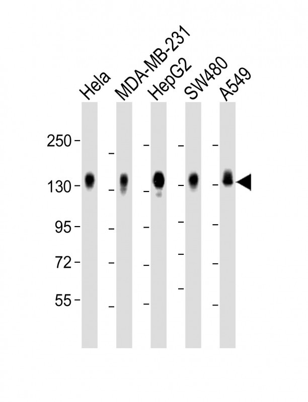
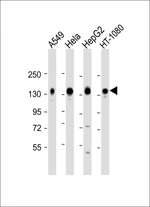
CD130 Antibody (C-term)
Affinity Purified Rabbit Polyclonal Antibody (Pab)


- SPECIFICATION
- CITATIONS
- PROTOCOLS
- BACKGROUND

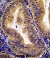
Application
| WB, IHC-P, E |
|---|---|
| Primary Accession | P40189 |
| Other Accession | NP_002175.2, NP_786943.1 |
| Reactivity | Human |
| Host | Rabbit |
| Clonality | Polyclonal |
| Isotype | Rabbit IgG |
| Calculated MW | 103537 Da |
| Antigen Region | 871-899 aa |
| Gene ID | 3572 |
|---|---|
| Other Names | Interleukin-6 receptor subunit beta, IL-6 receptor subunit beta, IL-6R subunit beta, IL-6R-beta, IL-6RB, CDw130, Interleukin-6 signal transducer, Membrane glycoprotein 130, gp130, Oncostatin-M receptor subunit alpha, CD130, IL6ST |
| Target/Specificity | This CD130 antibody is generated from rabbits immunized with a KLH conjugated synthetic peptide between 871-899 amino acids from the C-terminal region of human CD130. |
| Dilution | WB~~1:2000 IHC-P~~1:50~100 |
| Format | Purified polyclonal antibody supplied in PBS with 0.09% (W/V) sodium azide. This antibody is purified through a protein A column, followed by peptide affinity purification. |
| Storage | Maintain refrigerated at 2-8°C for up to 2 weeks. For long term storage store at -20°C in small aliquots to prevent freeze-thaw cycles. |
| Precautions | CD130 Antibody (C-term) is for research use only and not for use in diagnostic or therapeutic procedures. |
| Name | IL6ST (HGNC:6021) |
|---|---|
| Function | Signal-transducing molecule (PubMed:2261637). The receptor systems for IL6, LIF, OSM, CNTF, IL11, CTF1 and BSF3 can utilize IL6ST for initiating signal transmission. Binding of IL6 to IL6R induces IL6ST homodimerization and formation of a high-affinity receptor complex, which activates the intracellular JAK-MAPK and JAK-STAT3 signaling pathways (PubMed:2261637, PubMed:19915009, PubMed:23294003). That causes phosphorylation of IL6ST tyrosine residues which in turn activates STAT3 (PubMed:19915009, PubMed:23294003, PubMed:25731159). In parallel, the IL6 signaling pathway induces the expression of two cytokine receptor signaling inhibitors, SOCS1 and SOCS3, which inhibit JAK and terminate the activity of the IL6 signaling pathway as a negative feedback loop (By similarity). Also activates the yes- associated protein 1 (YAP) and NOTCH pathways to control inflammation- induced epithelial regeneration, independently of STAT3 (By similarity). Acts as a receptor for the neuroprotective peptide humanin as part of a complex with IL27RA/WSX1 and CNTFR (PubMed:19386761). Mediates signals which regulate immune response, hematopoiesis, pain control and bone metabolism (By similarity). Has a role in embryonic development (By similarity). Essential for survival of motor and sensory neurons and for differentiation of astrocytes (By similarity). Required for expression of TRPA1 in nociceptive neurons (By similarity). Required for the maintenance of PTH1R expression in the osteoblast lineage and for the stimulation of PTH-induced osteoblast differentiation (By similarity). Required for normal trabecular bone mass and cortical bone composition (By similarity). |
| Cellular Location | [Isoform 1]: Cell membrane; Single-pass type I membrane protein |
| Tissue Location | Found in all the tissues and cell lines examined (PubMed:2261637). Expression not restricted to IL6 responsive cells (PubMed:2261637). |

Thousands of laboratories across the world have published research that depended on the performance of antibodies from Abcepta to advance their research. Check out links to articles that cite our products in major peer-reviewed journals, organized by research category.
info@abcepta.com, and receive a free "I Love Antibodies" mug.

Provided below are standard protocols that you may find useful for product applications.
Background
The protein encoded by this gene is a signal transducer shared by many cytokines, including interleukin 6 (IL6), ciliary neurotrophic factor (CNTF), leukemia inhibitory factor (LIF), and oncostatin M (OSM). This protein functions as a part of the cytokine receptor complex. The activation of this protein is dependent upon the binding of cytokines to their receptors. vIL6, a protein related to IL6 and encoded by the Kaposi sarcoma-associated herpesvirus, can bypass the interleukin 6 receptor (IL6R) and directly activate this protein. Knockout studies in mice suggest that this gene plays a critical role in regulating myocyte apoptosis. Alternatively spliced transcript variants encoding distinct isoforms have been described. A related pseudogene has been identified on chromosome 17.
References
Bailey, S.D., et al. Diabetes Care 33(10):2250-2253(2010)
Goette, N.P., et al. Exp. Hematol. 38(10):868-876(2010)
Johnatty, S.E., et al. PLoS Genet. 6 (7), E1001016 (2010) :
Olsen, J.V., et al. Cell 127(3):635-648(2006)
Richards, P.J., et al. Arthritis Rheum. 54(5):1662-1672(2006)
If you have used an Abcepta product and would like to share how it has performed, please click on the "Submit Review" button and provide the requested information. Our staff will examine and post your review and contact you if needed.
If you have any additional inquiries please email technical services at tech@abcepta.com.
Ordering Information
Other Products
Shipping Information
