


 Foundational characteristics of cancer include proliferation, angiogenesis, migration, evasion of apoptosis, and cellular immortality. Find key markers for these cellular processes and antibodies to detect them.
 The SUMOplot™ Analysis Program predicts and scores sumoylation sites in your protein. SUMOylation is a post-translational modification involved in various cellular processes, such as nuclear-cytosolic transport, transcriptional regulation, apoptosis, protein stability, response to stress, and progression through the cell cycle.
 The Autophagy Receptor Motif Plotter predicts and scores autophagy receptor binding sites in your protein. Identifying proteins connected to this pathway is critical to understanding the role of autophagy in physiological as well as pathological processes such as development, differentiation, neurodegenerative diseases, stress, infection, and cancer.
MITF Antibody (N-term)
Affinity Purified Rabbit Polyclonal Antibody (Pab)
- SPECIFICATION
- CITATIONS: 1
- PROTOCOLS
- BACKGROUND

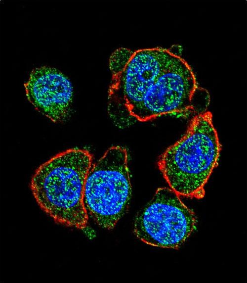
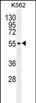
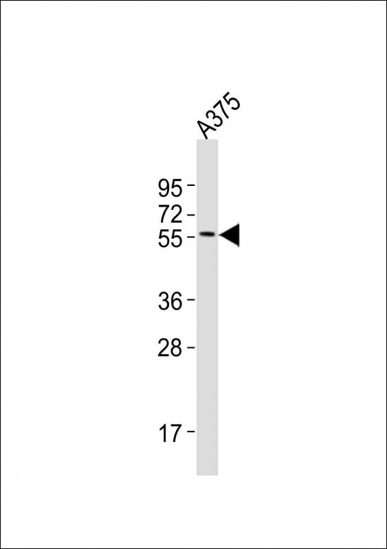
Application
| WB, IF, FC, E |
|---|---|
| Primary Accession | O75030 |
| Other Accession | Q08874, NP_937802.1, NP_001171896.1, O88368 |
| Reactivity | Human |
| Predicted | Mouse, Rat |
| Host | Rabbit |
| Clonality | Polyclonal |
| Isotype | Rabbit IgG |
| Calculated MW | 58795 Da |
| Antigen Region | 1-28 aa |
| Gene ID | 4286 |
|---|---|
| Other Names | Microphthalmia-associated transcription factor, Class E basic helix-loop-helix protein 32, bHLHe32, MITF, BHLHE32 |
| Target/Specificity | This MITF antibody is generated from rabbits immunized with a KLH conjugated synthetic peptide between 1-28 amino acids from the N-terminal region of human MITF. |
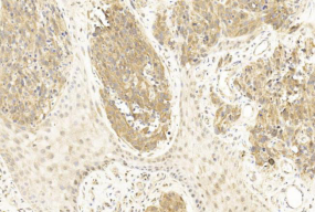
| Dilution | IF~~1:10~50 WB~~1:2000 IHC~~1:800 FC~~1:10~50 |
| Format | Purified polyclonal antibody supplied in PBS with 0.09% (W/V) sodium azide. This antibody is purified through a protein A column, followed by peptide affinity purification. |
| Storage | Maintain refrigerated at 2-8°C for up to 2 weeks. For long term storage store at -20°C in small aliquots to prevent freeze-thaw cycles. |
| Precautions | MITF Antibody (N-term) is for research use only and not for use in diagnostic or therapeutic procedures. |
| Name | MITF {ECO:0000303|PubMed:8069297, ECO:0000312|HGNC:HGNC:7105} |
|---|---|
| Function | Transcription factor that acts as a master regulator of melanocyte survival and differentiation as well as melanosome biogenesis (PubMed:10587587, PubMed:22647378, PubMed:27889061, PubMed:9647758). Binds to M-boxes (5'-TCATGTG-3') and symmetrical DNA sequences (E-boxes) (5'-CACGTG-3') found in the promoter of pigmentation genes, such as tyrosinase (TYR) (PubMed:10587587, PubMed:22647378, PubMed:27889061, PubMed:9647758). Involved in the cellular response to amino acid availability by acting downstream of MTOR: in the presence of nutrients, MITF phosphorylation by MTOR promotes its inactivation (PubMed:36608670). Upon starvation or lysosomal stress, inhibition of MTOR induces MITF dephosphorylation, resulting in transcription factor activity (PubMed:36608670). Plays an important role in melanocyte development by regulating the expression of tyrosinase (TYR) and tyrosinase-related protein 1 (TYRP1) (PubMed:10587587, PubMed:22647378, PubMed:27889061, PubMed:9647758). Plays a critical role in the differentiation of various cell types, such as neural crest-derived melanocytes, mast cells, osteoclasts and optic cup-derived retinal pigment epithelium (PubMed:10587587, PubMed:22647378, PubMed:27889061, PubMed:9647758). |
| Cellular Location | Nucleus. Cytoplasm. Lysosome membrane Note=When nutrients are present, recruited to the lysosomal membrane via association with GDP-bound RagC/RRAGC (or RagD/RRAGD): it is then phosphorylated by MTOR (PubMed:23401004, PubMed:36608670) Phosphorylation by MTOR promotes ubiquitination and degradation (PubMed:36608670). Conversely, inhibition of mTORC1, starvation and lysosomal disruption, promotes dephosphorylation and translocation to the nucleus (PubMed:36608670). Phosphorylation by MARK3/cTAK1 promotes association with 14-3-3/YWHA adapters and retention in the cytosol (PubMed:16822840). |
| Tissue Location | Expressed in melanocytes (at protein level). [Isoform C2]: Expressed in the kidney and retinal pigment epithelium. [Isoform H2]: Expressed in the kidney. [Isoform Mdel]: Expressed in melanocytes. |

Provided below are standard protocols that you may find useful for product applications.
Background
This gene encodes a transcription factor that contains both basic helix-loop-helix and leucine zipper structural features. It regulates the differentiation and development of melanocytes retinal pigment epithelium and is also responsible for pigment cell-specific transcription of the melanogenesis enzyme genes. Heterozygous mutations in the this gene cause auditory-pigmentary syndromes, such as Waardenburg syndrome type 2 and Tietz syndrome. Alternatively spliced transcript variants encoding different isoforms have been identified.
References
Wang, Y., et al. BMC Med 8, 14 (2010) : Shiohara, M., et al. Int J Lab Hematol 31(2):215-226(2009) Hershey, C.L., et al. Gene 347(1):73-82(2005) Miller, A.J., et al. J. Biol. Chem. 280(1):146-155(2005) Shibahara, S., et al. J. Investig. Dermatol. Symp. Proc. 6(1):99-104(2001)
If you have used an Abcepta product and would like to share how it has performed, please click on the "Submit Review" button and provide the requested information. Our staff will examine and post your review and contact you if needed.
If you have any additional inquiries please email technical services at tech@abcepta.com.

Ordering Information
Other Products
Shipping Information
