


 Foundational characteristics of cancer include proliferation, angiogenesis, migration, evasion of apoptosis, and cellular immortality. Find key markers for these cellular processes and antibodies to detect them.
 The SUMOplot™ Analysis Program predicts and scores sumoylation sites in your protein. SUMOylation is a post-translational modification involved in various cellular processes, such as nuclear-cytosolic transport, transcriptional regulation, apoptosis, protein stability, response to stress, and progression through the cell cycle.
 The Autophagy Receptor Motif Plotter predicts and scores autophagy receptor binding sites in your protein. Identifying proteins connected to this pathway is critical to understanding the role of autophagy in physiological as well as pathological processes such as development, differentiation, neurodegenerative diseases, stress, infection, and cancer.
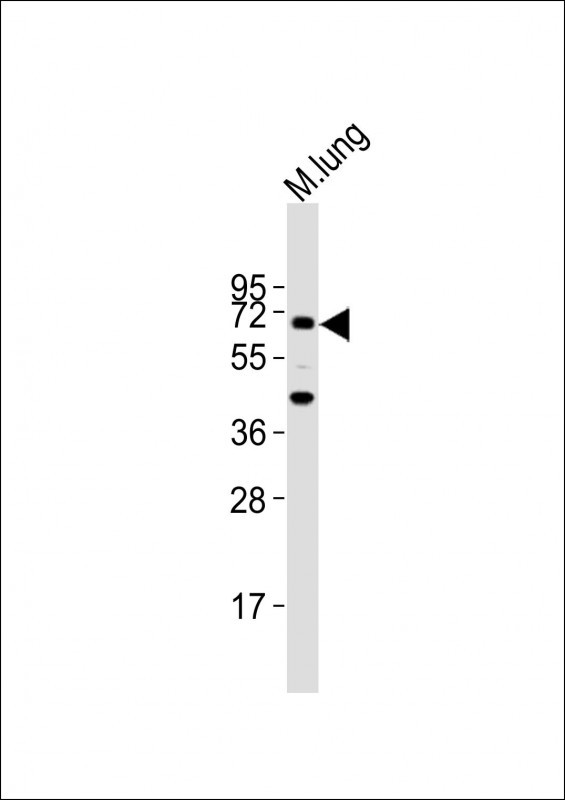
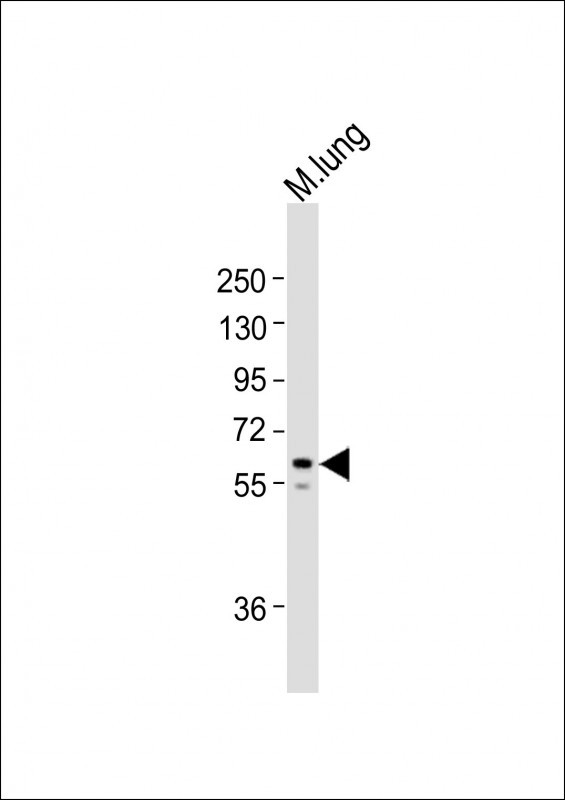
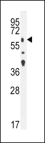
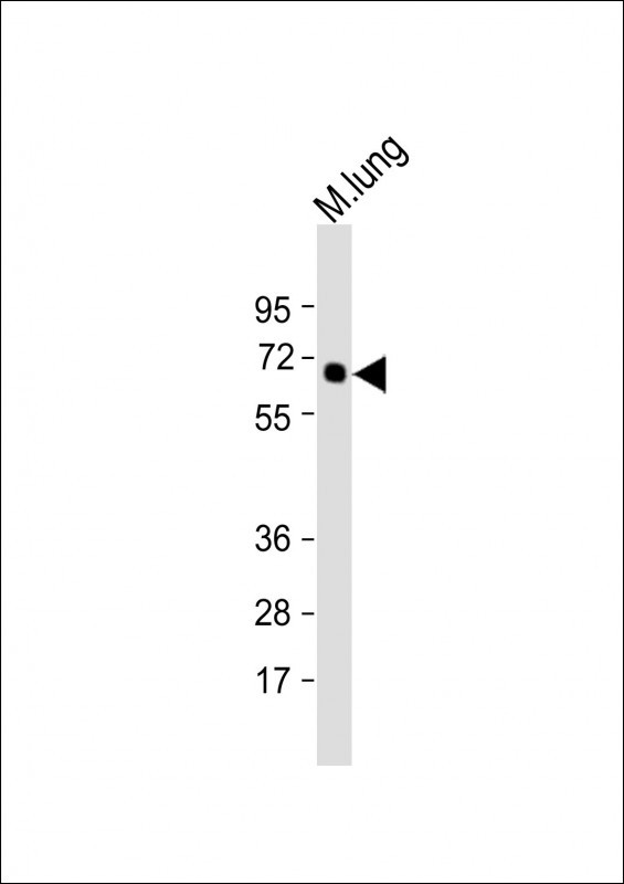
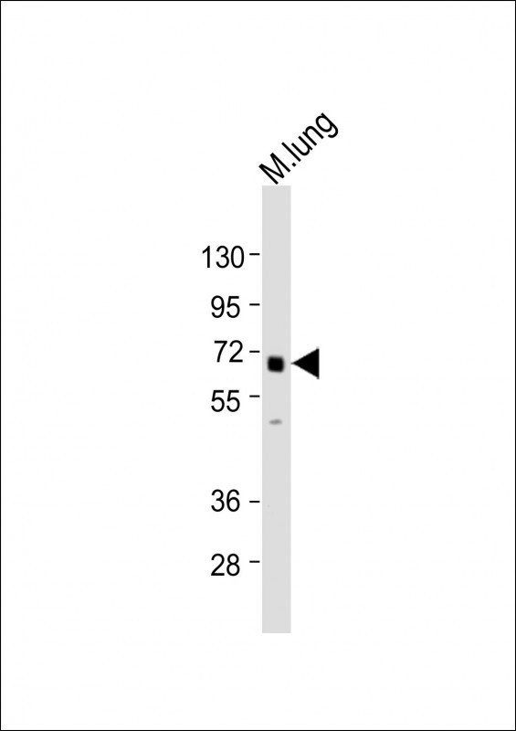
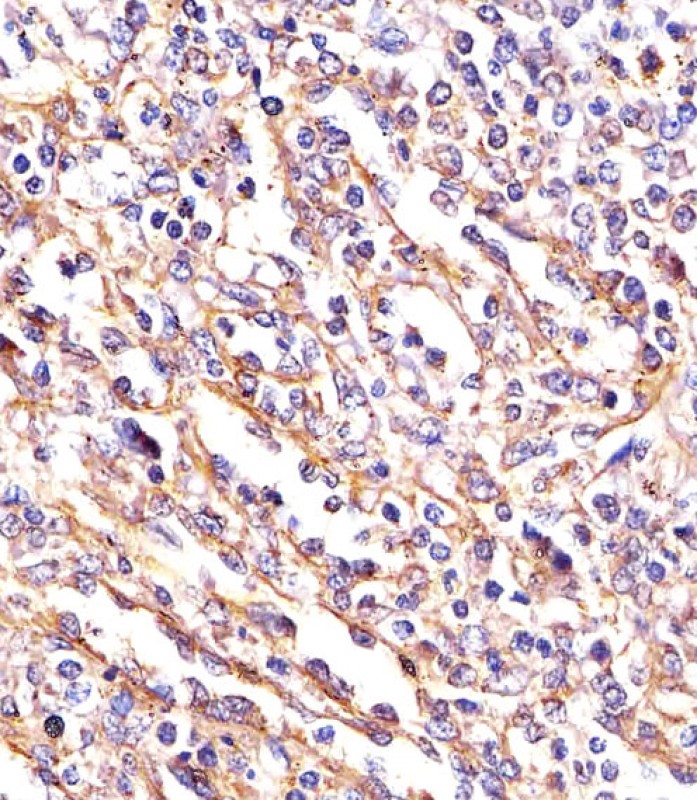
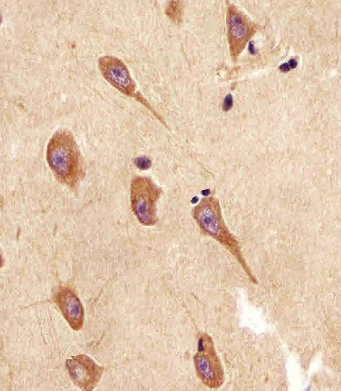
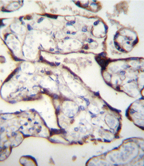
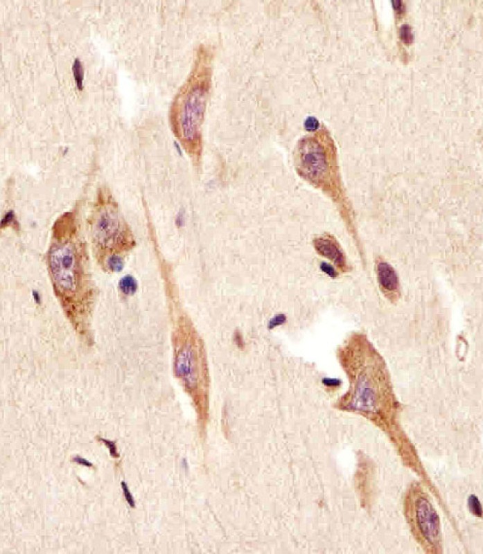
TGFBR2 Antibody (N-term)
Affinity Purified Rabbit Polyclonal Antibody (Pab)


- SPECIFICATION
- CITATIONS: 1
- PROTOCOLS
- BACKGROUND

Application
| WB, IHC-P, IF, E |
|---|---|
| Primary Accession | P37173 |
| Other Accession | NP_003233.4, NP_001020018.1 |
| Reactivity | Human, Mouse |
| Host | Rabbit |
| Clonality | Polyclonal |
| Isotype | Rabbit IgG |
| Calculated MW | 64568 Da |
| Antigen Region | 13-40 aa |
| Gene ID | 7048 |
|---|---|
| Other Names | TGF-beta receptor type-2, TGFR-2, TGF-beta type II receptor, Transforming growth factor-beta receptor type II, TGF-beta receptor type II, TbetaR-II, TGFBR2 |
| Target/Specificity | This TGFBR2 antibody is generated from rabbits immunized with a KLH conjugated synthetic peptide between 13-40 amino acids from the N-terminal region of human TGFBR2. |
| Dilution | IF~~1:10~50 WB~~1:1000 IHC-P~~1:25 |
| Format | Purified polyclonal antibody supplied in PBS with 0.09% (W/V) sodium azide. This antibody is purified through a protein A column, followed by peptide affinity purification. |
| Storage | Maintain refrigerated at 2-8°C for up to 2 weeks. For long term storage store at -20°C in small aliquots to prevent freeze-thaw cycles. |
| Precautions | TGFBR2 Antibody (N-term) is for research use only and not for use in diagnostic or therapeutic procedures. |
| Name | TGFBR2 |
|---|---|
| Function | Transmembrane serine/threonine kinase forming with the TGF- beta type I serine/threonine kinase receptor, TGFBR1, the non- promiscuous receptor for the TGF-beta cytokines TGFB1, TGFB2 and TGFB3. Transduces the TGFB1, TGFB2 and TGFB3 signal from the cell surface to the cytoplasm and thus regulates a plethora of physiological and pathological processes including cell cycle arrest in epithelial and hematopoietic cells, control of mesenchymal cell proliferation and differentiation, wound healing, extracellular matrix production, immunosuppression and carcinogenesis. The formation of the receptor complex composed of 2 TGFBR1 and 2 TGFBR2 molecules symmetrically bound to the cytokine dimer results in the phosphorylation and activation of TGFBR1 by the constitutively active TGFBR2. Activated TGFBR1 phosphorylates SMAD2 which dissociates from the receptor and interacts with SMAD4. The SMAD2-SMAD4 complex is subsequently translocated to the nucleus where it modulates the transcription of the TGF-beta-regulated genes. This constitutes the canonical SMAD-dependent TGF-beta signaling cascade. Also involved in non-canonical, SMAD-independent TGF-beta signaling pathways. |
| Cellular Location | Cell membrane; Single-pass type I membrane protein. Membrane raft |

Provided below are standard protocols that you may find useful for product applications.
Background
This gene encodes a member of the Ser/Thr protein kinase family and the TGFB receptor subfamily. The encoded protein is a transmembrane protein that has a protein kinase domain, forms a heterodimeric complex with another receptor protein, and binds TGF-beta. This receptor/ligand complex phosphorylates proteins, which then enter the nucleus and regulate the transcription of a subset of genes related to cell proliferation. Mutations in this gene have been associated with Marfan Syndrome, Loeys-Deitz Aortic Aneurysm Syndrome, and the development of various types of tumors. Alternatively spliced transcript variants encoding different isoforms have been characterized.
References
Inamoto, S., et al. Cardiovasc. Res. 88(3):520-529(2010)
Bianchini, G., et al. J. Clin. Oncol. 28(28):4316-4323(2010)
Bailey, S.D., et al. Diabetes Care 33(10):2250-2253(2010)
Kim, J.N., et al. Toxicology 275 (1-3), 29-35 (2010) :
Jugessur, A., et al. PLoS ONE 5 (7), E11493 (2010) :
If you have used an Abcepta product and would like to share how it has performed, please click on the "Submit Review" button and provide the requested information. Our staff will examine and post your review and contact you if needed.
If you have any additional inquiries please email technical services at tech@abcepta.com.
Ordering Information
Other Products
Shipping Information
