


 Foundational characteristics of cancer include proliferation, angiogenesis, migration, evasion of apoptosis, and cellular immortality. Find key markers for these cellular processes and antibodies to detect them.
 The SUMOplot™ Analysis Program predicts and scores sumoylation sites in your protein. SUMOylation is a post-translational modification involved in various cellular processes, such as nuclear-cytosolic transport, transcriptional regulation, apoptosis, protein stability, response to stress, and progression through the cell cycle.
 The Autophagy Receptor Motif Plotter predicts and scores autophagy receptor binding sites in your protein. Identifying proteins connected to this pathway is critical to understanding the role of autophagy in physiological as well as pathological processes such as development, differentiation, neurodegenerative diseases, stress, infection, and cancer.
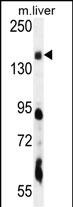
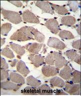
PIK3C2A Antibody (C-term)
Affinity Purified Rabbit Polyclonal Antibody (Pab)
- SPECIFICATION
- CITATIONS: 3
- PROTOCOLS
- BACKGROUND

Application
| IHC-P, WB, E |
|---|---|
| Primary Accession | O00443 |
| Other Accession | NP_002636.2 |
| Reactivity | Mouse |
| Host | Rabbit |
| Clonality | Polyclonal |
| Isotype | Rabbit IgG |
| Calculated MW | 190680 Da |
| Antigen Region | 1431-1458 aa |
| Gene ID | 5286 |
|---|---|
| Other Names | Phosphatidylinositol 4-phosphate 3-kinase C2 domain-containing subunit alpha, PI3K-C2-alpha, PtdIns-3-kinase C2 subunit alpha, Phosphoinositide 3-kinase-C2-alpha, PIK3C2A |
| Target/Specificity | This PIK3C2A antibody is generated from rabbits immunized with a KLH conjugated synthetic peptide between 1431-1458 amino acids from the C-terminal region of human PIK3C2A. |
| Dilution | WB~~1:1000 IHC-P~~1:10~50 |
| Format | Purified polyclonal antibody supplied in PBS with 0.09% (W/V) sodium azide. This antibody is purified through a protein A column, followed by peptide affinity purification. |
| Storage | Maintain refrigerated at 2-8°C for up to 2 weeks. For long term storage store at -20°C in small aliquots to prevent freeze-thaw cycles. |
| Precautions | PIK3C2A Antibody (C-term) is for research use only and not for use in diagnostic or therapeutic procedures. |
| Name | PIK3C2A |
|---|---|
| Function | Generates phosphatidylinositol 3-phosphate (PtdIns3P) and phosphatidylinositol 3,4-bisphosphate (PtdIns(3,4)P2) that act as second messengers. Has a role in several intracellular trafficking events. Functions in insulin signaling and secretion. Required for translocation of the glucose transporter SLC2A4/GLUT4 to the plasma membrane and glucose uptake in response to insulin-mediated RHOQ activation. Regulates insulin secretion through two different mechanisms: involved in glucose-induced insulin secretion downstream of insulin receptor in a pathway that involves AKT1 activation and TBC1D4/AS160 phosphorylation, and participates in the late step of insulin granule exocytosis probably in insulin granule fusion. Synthesizes PtdIns3P in response to insulin signaling. Functions in clathrin-coated endocytic vesicle formation and distribution. Regulates dynamin-independent endocytosis, probably by recruiting EEA1 to internalizing vesicles. In neurosecretory cells synthesizes PtdIns3P on large dense core vesicles. Participates in calcium induced contraction of vascular smooth muscle by regulating myosin light chain (MLC) phosphorylation through a mechanism involving Rho kinase-dependent phosphorylation of the MLCP-regulatory subunit MYPT1. May play a role in the EGF signaling cascade. May be involved in mitosis and UV-induced damage response. Required for maintenance of normal renal structure and function by supporting normal podocyte function. Involved in the regulation of ciliogenesis and trafficking of ciliary components (PubMed:31034465). |
| Cellular Location | Cell membrane. Cytoplasmic vesicle, clathrin-coated vesicle. Nucleus Cytoplasm Golgi apparatus, trans-Golgi network. Note=Inserts preferentially into membranes containing PtdIns(4,5)P2 (PubMed:17038310). Associated with RNA-containing structures (PubMed:11606566) |
| Tissue Location | Expressed in columnar and transitional epithelia, mononuclear cells, smooth muscle cells, and endothelial cells lining capillaries and small venules (at protein level). Ubiquitously expressed, with highest levels in heart, placenta and ovary, and lowest levels in the kidney. Detected at low levels in islets of Langerhans from type 2 diabetes mellitus individuals |

Provided below are standard protocols that you may find useful for product applications.
Background
The protein encoded by this gene belongs to the phosphoinositide 3-kinase (PI3K) family. PI3-kinases play roles in signaling pathways involved in cell proliferation, oncogenic transformation, cell survival, cell migration, and intracellular protein trafficking. This protein contains a lipid kinase catalytic domain as well as a C-terminal C2 domain, a characteristic of class II PI3-kinases. C2 domains act as calcium-dependent phospholipid binding motifs that mediate translocation of proteins to membranes, and may also mediate protein-protein interactions. The PI3-kinase activity of this protein is not sensitive to nanomolar levels of the inhibitor wortmanin. This protein was shown to be able to be activated by insulin and may be involved in integrin-dependent signaling.
References
Liu, C.Y., et al. Carcinogenesis 31(7):1259-1263(2010)
Rose, J.E., et al. Mol. Med. 16 (7-8), 247-253 (2010) :
Kostakis, G.C., et al. Oral Surg Oral Med Oral Pathol Oral Radiol Endod 109 (5), E53-E58 (2010) :
Koutros, S., et al. Cancer Res. 70(6):2389-2396(2010)
Ng, S.K., et al. Biochem. Biophys. Res. Commun. 387(2):310-315(2009)
If you have used an Abcepta product and would like to share how it has performed, please click on the "Submit Review" button and provide the requested information. Our staff will examine and post your review and contact you if needed.
If you have any additional inquiries please email technical services at tech@abcepta.com.
Ordering Information
Other Products
Shipping Information
