


 Foundational characteristics of cancer include proliferation, angiogenesis, migration, evasion of apoptosis, and cellular immortality. Find key markers for these cellular processes and antibodies to detect them.
 The SUMOplot™ Analysis Program predicts and scores sumoylation sites in your protein. SUMOylation is a post-translational modification involved in various cellular processes, such as nuclear-cytosolic transport, transcriptional regulation, apoptosis, protein stability, response to stress, and progression through the cell cycle.
 The Autophagy Receptor Motif Plotter predicts and scores autophagy receptor binding sites in your protein. Identifying proteins connected to this pathway is critical to understanding the role of autophagy in physiological as well as pathological processes such as development, differentiation, neurodegenerative diseases, stress, infection, and cancer.
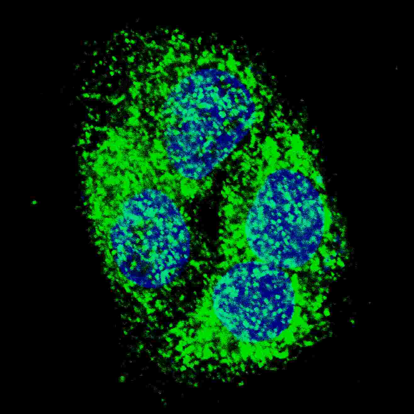
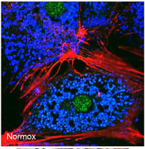
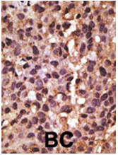
BNIP3 Antibody (BH3 Domain Specific)
Purified Rabbit Polyclonal Antibody (Pab)
- SPECIFICATION
- CITATIONS: 3
- PROTOCOLS
- BACKGROUND

Application
| IHC-P, IF, E |
|---|---|
| Primary Accession | Q12983 |
| Reactivity | Human, Mouse |
| Host | Rabbit |
| Clonality | Polyclonal |
| Isotype | Rabbit IgG |
| Calculated MW | 21541 Da |
| Antigen Region | 215-252 aa |
| Gene ID | 664 |
|---|---|
| Other Names | BCL2/adenovirus E1B 19 kDa protein-interacting protein 3, BNIP3, NIP3 |
| Target/Specificity | This BNIP3 antibody is generated from rabbits immunized with a KLH conjugated synthetic peptide between 215-252 amino acids from human BNIP3. |
| Dilution | IF~~1:50~100 IHC-P~~1:50~100 |
| Format | Purified polyclonal antibody supplied in PBS with 0.09% (W/V) sodium azide. This antibody is prepared by Saturated Ammonium Sulfate (SAS) precipitation followed by dialysis against PBS. |
| Storage | Maintain refrigerated at 2-8°C for up to 2 weeks. For long term storage store at -20°C in small aliquots to prevent freeze-thaw cycles. |
| Precautions | BNIP3 Antibody (BH3 Domain Specific) is for research use only and not for use in diagnostic or therapeutic procedures. |
| Name | BNIP3 (HGNC:1084) |
|---|---|
| Synonyms | NIP3 |
| Function | Apoptosis-inducing protein that can overcome BCL2 suppression. May play a role in repartitioning calcium between the two major intracellular calcium stores in association with BCL2. Involved in mitochondrial quality control via its interaction with SPATA18/MIEAP: in response to mitochondrial damage, participates in mitochondrial protein catabolic process (also named MALM) leading to the degradation of damaged proteins inside mitochondria. The physical interaction of SPATA18/MIEAP, BNIP3 and BNIP3L/NIX at the mitochondrial outer membrane regulates the opening of a pore in the mitochondrial double membrane in order to mediate the translocation of lysosomal proteins from the cytoplasm to the mitochondrial matrix. Plays an important role in the calprotectin (S100A8/A9)-induced cell death pathway. |
| Cellular Location | Mitochondrion. Mitochondrion outer membrane; Single-pass membrane protein. Note=Coexpression with the EIB 19-kDa protein results in a shift in NIP3 localization pattern to the nuclear envelope. Colocalizes with ACAA2 in the mitochondria. Colocalizes with SPATA18 at the mitochondrion outer membrane |

Provided below are standard protocols that you may find useful for product applications.
Background
NIP3 is a member of the BCL2/adenovirus E1B 19 kd-interacting protein (BNIP) family. It interacts with the E1B 19 kDa protein which is responsible for the protection of virally-induced cell death, as well as E1B 19 kDa-like sequences of BCL2, also an apoptotic protector. NIP3 contains a BH3 domain and a transmembrane domain, which have been associated with pro-apoptotic function. The dimeric mitochondrial protein is known to induce apoptosis, even in the presence of BCL2.
References
References for protein:
1.Kothari, S., et al., Oncogene 22(30):4734-4744 (2003).
2.Lee, S.M., et al., Life Sci. 71(19):2267-2277 (2002).
3.Ray, R., et al., J. Biol. Chem. 275(2):1439-1448 (2000).
4.Chen, G., et al., J. Biol. Chem. 274(1):7-10 (1999).
5.Yasuda, M., et al., J. Biol. Chem. 273(20):12415-12421 (1998).
References for HepG2 cell line:
1. Knowles BB, et al. (1980). Human hepatocellular carcinoma cell lines secrete the major plasma proteins and hepatitis B surface antigen. Science 209: 497-499.[ PubMed: 6248960].
2. Darlington GJ, et al. (1987). Growth and hepatospecific gene expression of human hepatoma cells in a defined medium. In Vitro Cell. Dev. Biol. 23: 349-354.[PubMed: 3034851].
3. Ihrke, G; Neufeld, EB; Meads, T; Shanks, MR; Cassio, D; Laurent, M; Schroer, TA; Pagano, RE et al. (1993). "WIF-B cells: an in vitro model for studies of hepatocyte polarity". Journal of Cell Biology 123 (6): 1761–1775. [PubMed:7506266].
4. Mersch-Sundermann, V.; Knasmüller, S.; Wu, X. J.; Darroudi, F.; Kassie, F. (2004). "Use of a human-derived liver cell line for the detection of cytoprotective, antigenotoxic and cogenotoxic agents". Toxicology 198 (1–3): 329–340. [PubMed:15138059].
If you have used an Abcepta product and would like to share how it has performed, please click on the "Submit Review" button and provide the requested information. Our staff will examine and post your review and contact you if needed.
If you have any additional inquiries please email technical services at tech@abcepta.com.
Ordering Information
Other Products
Shipping Information
