


 Foundational characteristics of cancer include proliferation, angiogenesis, migration, evasion of apoptosis, and cellular immortality. Find key markers for these cellular processes and antibodies to detect them.
 The SUMOplot™ Analysis Program predicts and scores sumoylation sites in your protein. SUMOylation is a post-translational modification involved in various cellular processes, such as nuclear-cytosolic transport, transcriptional regulation, apoptosis, protein stability, response to stress, and progression through the cell cycle.
 The Autophagy Receptor Motif Plotter predicts and scores autophagy receptor binding sites in your protein. Identifying proteins connected to this pathway is critical to understanding the role of autophagy in physiological as well as pathological processes such as development, differentiation, neurodegenerative diseases, stress, infection, and cancer.
Mouse TLR1 Antibody (C-term)
Purified Rabbit Polyclonal Antibody (Pab)
- SPECIFICATION
- CITATIONS
- PROTOCOLS
- BACKGROUND

Application
| IHC-P, WB, E |
|---|---|
| Primary Accession | Q9EPQ1 |
| Reactivity | Mouse |
| Host | Rabbit |
| Clonality | Polyclonal |
| Isotype | Rabbit IgG |
| Calculated MW | 90673 Da |
| Antigen Region | 764-795 aa |
| Gene ID | 21897 |
|---|---|
| Other Names | Toll-like receptor 1, Toll/interleukin-1 receptor-like protein, TIL, CD281, Tlr1 |
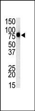
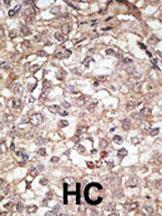
| Target/Specificity | This Mouse TLR1 antibody is generated from rabbits immunized with a KLH conjugated synthetic peptide between 764-795 amino acids from the C-terminal region of mouse TLR1. |
| Dilution | WB~~1:1000 IHC-P~~1:50~100 |
| Format | Purified polyclonal antibody supplied in PBS with 0.09% (W/V) sodium azide. This antibody is purified through a protein A column, followed by peptide affinity purification. |
| Storage | Maintain refrigerated at 2-8°C for up to 2 weeks. For long term storage store at -20°C in small aliquots to prevent freeze-thaw cycles. |
| Precautions | Mouse TLR1 Antibody (C-term) is for research use only and not for use in diagnostic or therapeutic procedures. |
| Name | Tlr1 |
|---|---|
| Function | Participates in the innate immune response to microbial agents. Specifically recognizes diacylated and triacylated lipopeptides. Cooperates with TLR2 to mediate the innate immune response to bacterial lipoproteins or lipopeptides. Forms the activation cluster TLR2:TLR1:CD14 in response to triacylated lipopeptides, this cluster triggers signaling from the cell surface and subsequently is targeted to the Golgi in a lipid-raft dependent pathway. Acts via MYD88 and TRAF6, leading to NF-kappa-B activation, cytokine secretion and the inflammatory response (By similarity). Acts as a coreceptor for M.tuberculosis lipoproteins LprG, LpqH and PhoS1 (pstS1), in conjunction with TLR2 and for some but not all lipoproteins CD14 and/or CD36. The lipoproteins act as agonists to modulate antigen presenting cell functions in response to the pathogen (PubMed:19362712). |
| Cellular Location | Cell membrane; Single-pass type I membrane protein. Cytoplasmic vesicle, phagosome membrane; Single-pass type I membrane protein. Membrane raft {ECO:0000250|UniProtKB:Q15399}. Golgi apparatus {ECO:0000250|UniProtKB:Q15399}. Note=Does not reside in lipid rafts before stimulation but accumulates increasingly in the raft upon the presence of the microbial ligand. In response to triacylated lipoproteins, TLR2:TLR1 heterodimers are recruited in lipid rafts, this recruitment determine the intracellular targeting to the Golgi apparatus. {ECO:0000250|UniProtKB:Q15399} |

Thousands of laboratories across the world have published research that depended on the performance of antibodies from Abcepta to advance their research. Check out links to articles that cite our products in major peer-reviewed journals, organized by research category.
info@abcepta.com, and receive a free "I Love Antibodies" mug.

Provided below are standard protocols that you may find useful for product applications.
Background
Higher animals establish host defense by orchestrating innate and adaptive immunity. This is mediated by professional antigen presenting cells, i.e. dendritic cells (DCs). DCs can incorporate pathogens, produce a variety of cytokines, maturate, and present pathogen-derived peptides to T cells, thereby inducing T cell activation and differentiation. These responses are triggered by microbial recognition through type I transmembrane proteins, Toll-like receptors (TLRs) on DCs. TLRs consist of ten members and each TLR is involved in recognizing a variety of microorganism-derived molecular structures. TLR ligands include cell wall components, proteins, nucleic acids, and synthetic chemical compounds, all of which can activate DCs as immune adjuvants. Each TLR can activate DCs in a similar, but distinct manner. For example, TLRs can be divided into subgroups according to their type I interferon (IFN) inducing ability. TLR2 cannot induce IFN-alpha or IFN-beta, but TLR4 can lead to IFN-beta production. Meanwhile, TLR3, TLR7, and TLR9 can induce both IFN-alpha and IFN-beta. Recent evidences suggest that cytoplamic adapters for TLRs are especially crucial for this functional heterogeneity.
References
Hajjar, A.M., et al., J. Immunol. 166(1):15-19 (2001).
Ozinsky, A., et al., Proc. Natl. Acad. Sci. U.S.A. 97(25):13766-13771 (2000).
If you have used an Abcepta product and would like to share how it has performed, please click on the "Submit Review" button and provide the requested information. Our staff will examine and post your review and contact you if needed.
If you have any additional inquiries please email technical services at tech@abcepta.com.
Ordering Information
Other Products
Shipping Information
