


 Foundational characteristics of cancer include proliferation, angiogenesis, migration, evasion of apoptosis, and cellular immortality. Find key markers for these cellular processes and antibodies to detect them.
 The SUMOplot™ Analysis Program predicts and scores sumoylation sites in your protein. SUMOylation is a post-translational modification involved in various cellular processes, such as nuclear-cytosolic transport, transcriptional regulation, apoptosis, protein stability, response to stress, and progression through the cell cycle.
 The Autophagy Receptor Motif Plotter predicts and scores autophagy receptor binding sites in your protein. Identifying proteins connected to this pathway is critical to understanding the role of autophagy in physiological as well as pathological processes such as development, differentiation, neurodegenerative diseases, stress, infection, and cancer.
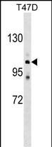
ATP2C1 Antibody (C-term)
Affinity Purified Rabbit Polyclonal Antibody (Pab)
- SPECIFICATION
- CITATIONS
- PROTOCOLS
- BACKGROUND

Application
| WB, E |
|---|---|
| Primary Accession | P98194 |
| Other Accession | NP_001001486.1, NP_001001485.1 |
| Reactivity | Human |
| Host | Rabbit |
| Clonality | Polyclonal |
| Isotype | Rabbit IgG |
| Calculated MW | 100577 Da |
| Antigen Region | 882-909 aa |
| Gene ID | 27032 |
|---|---|
| Other Names | Calcium-transporting ATPase type 2C member 1, ATPase 2C1, ATP-dependent Ca(2+) pump PMR1, ATP2C1, KIAA1347, PMR1L |
| Target/Specificity | This ATP2C1 antibody is generated from rabbits immunized with a KLH conjugated synthetic peptide between 882-909 amino acids from the C-terminal region of human ATP2C1. |
| Dilution | WB~~1:1000 |
| Format | Purified polyclonal antibody supplied in PBS with 0.09% (W/V) sodium azide. This antibody is purified through a protein A column, followed by peptide affinity purification. |
| Storage | Maintain refrigerated at 2-8°C for up to 2 weeks. For long term storage store at -20°C in small aliquots to prevent freeze-thaw cycles. |
| Precautions | ATP2C1 Antibody (C-term) is for research use only and not for use in diagnostic or therapeutic procedures. |
| Name | ATP2C1 {ECO:0000303|PubMed:10615129, ECO:0000312|HGNC:HGNC:13211} |
|---|---|
| Function | ATP-driven pump that supplies the Golgi apparatus with Ca(2+) and Mn(2+) ions, both essential cofactors for processing and trafficking of newly synthesized proteins in the secretory pathway (PubMed:16192278, PubMed:30923126, PubMed:21187401, PubMed:12707275, PubMed:20439740). Within a catalytic cycle, acquires Ca(2+) or Mn(2+) ions on the cytoplasmic side of the membrane and delivers them to the lumenal side. The transfer of ions across the membrane is coupled to ATP hydrolysis and is associated with a transient phosphorylation that shifts the pump conformation from inward-facing to outward-facing state (PubMed:16192278, PubMed:16332677, PubMed:30923126). Plays a primary role in the maintenance of Ca(2+) homeostasis in the trans-Golgi compartment with a functional impact on Golgi and post-Golgi protein sorting as well as a structural impact on cisternae morphology (PubMed:20439740, PubMed:14632183). Responsible for loading the Golgi stores with Ca(2+) ions in keratinocytes, contributing to keratinocyte differentiation and epidermis integrity (PubMed:14632183, PubMed:10615129, PubMed:20439740). Participates in Ca(2+) and Mn(2+) ions uptake into the Golgi store of hippocampal neurons and regulates protein trafficking required for neural polarity (By similarity). May also play a role in the maintenance of Ca(2+) and Mn(2+) homeostasis and signaling in the cytosol while preventing cytotoxicity (PubMed:21187401). |
| Cellular Location | Golgi apparatus, trans-Golgi network membrane; Multi-pass membrane protein. Golgi apparatus, Golgi stack membrane; Multi-pass membrane protein. Note=During neuron differentiation, shifts from juxtanuclear Golgi position to multiple Golgi structures distributed over the neural soma with a predominance in the apical dendritic trunk {ECO:0000250|UniProtKB:Q80XR2} |
| Tissue Location | Found in most tissues except colon, thymus, spleen and leukocytes (PubMed:15831496). Expressed in keratinocytes (at protein level) (PubMed:15831496, PubMed:14632183) |

Thousands of laboratories across the world have published research that depended on the performance of antibodies from Abcepta to advance their research. Check out links to articles that cite our products in major peer-reviewed journals, organized by research category.
info@abcepta.com, and receive a free "I Love Antibodies" mug.

Provided below are standard protocols that you may find useful for product applications.
Background
The protein encoded by this gene belongs to the family of P-type cation transport ATPases. This magnesium-dependent enzyme catalyzes the hydrolysis of ATP coupled with the transport of the calcium. Defects in this gene cause Hailey-Hailey disease, an autosomal dominant disorder. Alternatively spliced transcript variants encoding different isoforms have been identified.
References
Baron, S., et al. Biochim. Biophys. Acta 1798(8):1512-1521(2010)
Davila, S., et al. Genes Immun. 11(3):232-238(2010)
Tian, H., et al. J. Dermatol. Sci. 58(1):80-82(2010)
Ding, Y.G., et al. Clin. Exp. Dermatol. 34 (8), E968-E971 (2009) :
Nechama, M., et al. BMC Cell Biol. 10, 70 (2009) :
If you have used an Abcepta product and would like to share how it has performed, please click on the "Submit Review" button and provide the requested information. Our staff will examine and post your review and contact you if needed.
If you have any additional inquiries please email technical services at tech@abcepta.com.

Ordering Information
Other Products
Shipping Information
