


 Foundational characteristics of cancer include proliferation, angiogenesis, migration, evasion of apoptosis, and cellular immortality. Find key markers for these cellular processes and antibodies to detect them.
 The SUMOplot™ Analysis Program predicts and scores sumoylation sites in your protein. SUMOylation is a post-translational modification involved in various cellular processes, such as nuclear-cytosolic transport, transcriptional regulation, apoptosis, protein stability, response to stress, and progression through the cell cycle.
 The Autophagy Receptor Motif Plotter predicts and scores autophagy receptor binding sites in your protein. Identifying proteins connected to this pathway is critical to understanding the role of autophagy in physiological as well as pathological processes such as development, differentiation, neurodegenerative diseases, stress, infection, and cancer.
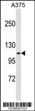
FARP1 Antibody (N-term)
Affinity Purified Rabbit Polyclonal Antibody (Pab)
- SPECIFICATION
- CITATIONS
- PROTOCOLS
- BACKGROUND

Application
| WB, E |
|---|---|
| Primary Accession | Q9Y4F1 |
| Other Accession | F1P065, NP_005757.1 |
| Reactivity | Human |
| Predicted | Chicken |
| Host | Rabbit |
| Clonality | Polyclonal |
| Isotype | Rabbit IgG |
| Calculated MW | 118633 Da |
| Antigen Region | 62-90 aa |
| Gene ID | 10160 |
|---|---|
| Other Names | FERM, RhoGEF and pleckstrin domain-containing protein 1, Chondrocyte-derived ezrin-like protein, Pleckstrin homology domain-containing family C member 2, PH domain-containing family C member 2, FARP1, CDEP, PLEKHC2 |
| Target/Specificity | This FARP1 antibody is generated from rabbits immunized with a KLH conjugated synthetic peptide between 62-90 amino acids from the N-terminal region of human FARP1. |
| Dilution | WB~~1:1000 |
| Format | Purified polyclonal antibody supplied in PBS with 0.09% (W/V) sodium azide. This antibody is purified through a protein A column, followed by peptide affinity purification. |
| Storage | Maintain refrigerated at 2-8°C for up to 2 weeks. For long term storage store at -20°C in small aliquots to prevent freeze-thaw cycles. |
| Precautions | FARP1 Antibody (N-term) is for research use only and not for use in diagnostic or therapeutic procedures. |
| Name | FARP1 |
|---|---|
| Synonyms | CDEP, PLEKHC2 |
| Function | Functions as a guanine nucleotide exchange factor for RAC1. May play a role in semaphorin signaling. Plays a role in the assembly and disassembly of dendritic filopodia, the formation of dendritic spines, regulation of dendrite length and ultimately the formation of synapses (By similarity). |
| Cellular Location | Cell membrane; Peripheral membrane protein; Cytoplasmic side. Synapse. Synapse, synaptosome Cytoplasm, cytosol. Cell projection, filopodium. Cell projection, dendrite. Cell projection, dendritic spine. Note=Recruited to the cell membrane via interaction with CADM1. |
| Tissue Location | Detected in cAMP-treated chondrocytes, but not in untreated chondrocytes. Detected in fetal brain, heart and spleen, and in adult testis, kidney and lung. |

Thousands of laboratories across the world have published research that depended on the performance of antibodies from Abcepta to advance their research. Check out links to articles that cite our products in major peer-reviewed journals, organized by research category.
info@abcepta.com, and receive a free "I Love Antibodies" mug.

Provided below are standard protocols that you may find useful for product applications.
Background
This gene was originally isolated through subtractive hybridization due to its increased expression in differentiated chondrocytes versus dedifferentiated chondrocytes. The resulting protein contains a predicted ezrin-like domain, a Dbl homology domain, and a pleckstrin homology domain. It is believed to be a member of the band 4.1 superfamily whose members link the cytoskeleton to the cell membrane. Two alternatively spliced transcript variants encoding distinct isoforms have been found for this gene.
References
Stein, J.L., et al. Neuroimage 53(3):1160-1174(2010)
Rose, J.E., et al. Mol. Med. 16 (7-8), 247-253 (2010) :
Evangelou, E., et al. Am. J. Med. Genet. B Neuropsychiatr. Genet. 153B (1), 220-228 (2010) :
Sugiyama, N., et al. Mol. Cell Proteomics 6(6):1103-1109(2007)
Olsen, J.V., et al. Cell 127(3):635-648(2006)
If you have used an Abcepta product and would like to share how it has performed, please click on the "Submit Review" button and provide the requested information. Our staff will examine and post your review and contact you if needed.
If you have any additional inquiries please email technical services at tech@abcepta.com.
Ordering Information
Other Products
Shipping Information
