


 Foundational characteristics of cancer include proliferation, angiogenesis, migration, evasion of apoptosis, and cellular immortality. Find key markers for these cellular processes and antibodies to detect them.
 The SUMOplot™ Analysis Program predicts and scores sumoylation sites in your protein. SUMOylation is a post-translational modification involved in various cellular processes, such as nuclear-cytosolic transport, transcriptional regulation, apoptosis, protein stability, response to stress, and progression through the cell cycle.
 The Autophagy Receptor Motif Plotter predicts and scores autophagy receptor binding sites in your protein. Identifying proteins connected to this pathway is critical to understanding the role of autophagy in physiological as well as pathological processes such as development, differentiation, neurodegenerative diseases, stress, infection, and cancer.
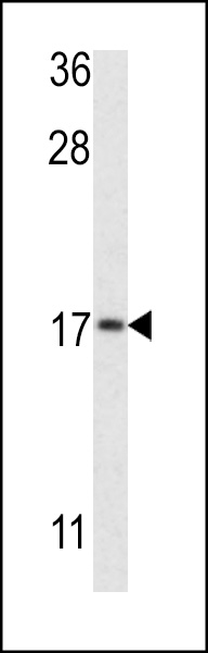
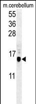
APG8b (MAP1LC3B)-T93/Y99 Antibody (Center)
Affinity Purified Rabbit Polyclonal Antibody (Pab)
- SPECIFICATION
- CITATIONS: 1
- PROTOCOLS
- BACKGROUND

Application
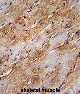
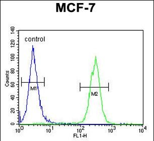
| FC, IHC-P, WB, E |
|---|---|
| Primary Accession | Q9GZQ8 |
| Other Accession | A6NCE7, NP_073729.1 |
| Reactivity | Human, Mouse |
| Host | Rabbit |
| Clonality | Polyclonal |
| Isotype | Rabbit IgG |
| Calculated MW | 14688 Da |
| Antigen Region | 74-106 aa |
| Gene ID | 81631 |
|---|---|
| Other Names | Microtubule-associated proteins 1A/1B light chain 3B, Autophagy-related protein LC3 B, Autophagy-related ubiquitin-like modifier LC3 B, MAP1 light chain 3-like protein 2, MAP1A/MAP1B light chain 3 B, MAP1A/MAP1B LC3 B, Microtubule-associated protein 1 light chain 3 beta, MAP1LC3B, MAP1ALC3 |
| Target/Specificity | This APG8b (MAP1LC3B)-T93/Y99 antibody is generated from rabbits immunized with a KLH conjugated synthetic peptide between 74-106 amino acids from the Central region of human APG8b (MAP1LC3B)-T93/Y99. |
| Dilution | WB~~1:1000 IHC-P~~1:50~100 FC~~1:10~50 |
| Format | Purified polyclonal antibody supplied in PBS with 0.09% (W/V) sodium azide. This antibody is purified through a protein A column, followed by peptide affinity purification. |
| Storage | Maintain refrigerated at 2-8°C for up to 2 weeks. For long term storage store at -20°C in small aliquots to prevent freeze-thaw cycles. |
| Precautions | APG8b (MAP1LC3B)-T93/Y99 Antibody (Center) is for research use only and not for use in diagnostic or therapeutic procedures. |
| Name | MAP1LC3B (HGNC:13352) |
|---|---|
| Synonyms | MAP1ALC3 |
| Function | Ubiquitin-like modifier involved in formation of autophagosomal vacuoles (autophagosomes) (PubMed:20418806, PubMed:23209295, PubMed:28017329). Plays a role in mitophagy which contributes to regulate mitochondrial quantity and quality by eliminating the mitochondria to a basal level to fulfill cellular energy requirements and preventing excess ROS production (PubMed:23209295, PubMed:28017329). In response to cellular stress and upon mitochondria fission, binds C-18 ceramides and anchors autophagolysosomes to outer mitochondrial membranes to eliminate damaged mitochondria (PubMed:22922758). While LC3s are involved in elongation of the phagophore membrane, the GABARAP/GATE-16 subfamily is essential for a later stage in autophagosome maturation (PubMed:20418806, PubMed:23209295, PubMed:28017329). Promotes primary ciliogenesis by removing OFD1 from centriolar satellites via the autophagic pathway (PubMed:24089205). Through its interaction with the reticulophagy receptor TEX264, participates in the remodeling of subdomains of the endoplasmic reticulum into autophagosomes upon nutrient stress, which then fuse with lysosomes for endoplasmic reticulum turnover (PubMed:31006537, PubMed:31006538). Upon nutrient stress, directly recruits cofactor JMY to the phagophore membrane surfaces and promotes JMY's actin nucleation activity and autophagosome biogenesis during autophagy (PubMed:30420355). |
| Cellular Location | Cytoplasmic vesicle, autophagosome membrane; Lipid-anchor Endomembrane system; Lipid-anchor Mitochondrion membrane; Lipid-anchor. Cytoplasm, cytoskeleton {ECO:0000250|UniProtKB:Q9CQV6}. Cytoplasmic vesicle. Note=LC3-II binds to the autophagic membranes. LC3-II localizes with the mitochondrial inner membrane during Parkin-mediated mitophagy (PubMed:28017329). Localizes also to discrete punctae along the ciliary axoneme |
| Tissue Location | Most abundant in heart, brain, skeletal muscle and testis. Little expression observed in liver |

Provided below are standard protocols that you may find useful for product applications.
Background
The product of this gene is a subunit of neuronal microtubule-associated MAP1A and MAP1B proteins, which are involved in microtubule assembly and important for neurogenesis. Studies on the rat homolog implicate a role for this gene in autophagy, a process that involves the bulk degradation of cytoplasmic component.
References
Rouschop, K.M., et al. J. Clin. Invest. 120(1):127-141(2010)
Othman, E.Q., et al. J. Clin. Lab. Anal. 23(4):249-258(2009)
Komatsu, M., et al. Cell 131(6):1149-1163(2007)
Stelzl, U., et al. Cell 122(6):957-968(2005)
Tanida, I., et al. J. Biol. Chem. 279(46):47704-47710(2004)
He, H., et al. J. Biol. Chem. 278(31):29278-29287(2003)
Tanida, I., et al. J. Biol. Chem. 277(16):13739-13744(2002)
Tanida, I., et al. Biochem. Biophys. Res. Commun. 292(1):256-262(2002)
Kabeya, Y., et al. EMBO J. 19(21):5720-5728(2000)
Simpson, J.C., et al. EMBO Rep. 1(3):287-292(2000)
If you have used an Abcepta product and would like to share how it has performed, please click on the "Submit Review" button and provide the requested information. Our staff will examine and post your review and contact you if needed.
If you have any additional inquiries please email technical services at tech@abcepta.com.
Ordering Information
Other Products
Shipping Information
